The cerebral cortex in humans reflects the pinnacle of phylogenetic development with respect to cortical size and complexity. It is estimated that the human cerebral cortex contains 15 billion neurons, encompassing an area of approximately 220 cm2. Much of the cortex is out of view because of the convexities of the sulci. Different parts of the cerebral cortex receive many afferent fibers. The largest source of fibers is from the thalamus, but other regions, such as the brainstem monoaminergic systems and parts of the basal forebrain, contribute fibers as well. Cortical afferent fibers also arise from other regions of the cortex (association fibers) and from the contralateral cerebral cortex (via the corpus callosum). The complexities of the functions attributable to the cerebral cortex derive, in part, from the nature of the histological organization of the cortex, which differs from one cortical region to another. The histological arrangement of a given region of cortex can be subdivided into basic functional units called cortical columns. Collectively, the integrated actions of groups of cortical columns serve to mediate voluntary motor activity, sensory perception, learning, memory, language functions, and affective processes.
The varied processes associated with the cerebral cortex can be better understood by examining the nature of the organization of different regions of cortex, including the functional properties of cortical columns, followed by an analysis of the afferent and efferent connections of this region of the brain. Accordingly, this topic begins with an examination of the histology of the cerebral cortex followed by a review and discussion of the input-output relationships of the cerebral cortex.
Anatomical and Functional Characteristics of the Gray Matter of the Cerebral Cortex
We begin this topic by considering the organization of the basic functional units of the cerebral cortex. The functional units are composed of narrow bands of gray matter of cortex arranged vertically through all cell layers called cortical columns. To understand how a cortical column functions, as well as its relationship to the overall functions of the region of cortex of which it is a part, it is first necessary to consider the histological arrangement of the gray matter of the cerebral cortex, which differs somewhat from region to region.
Morphological Features
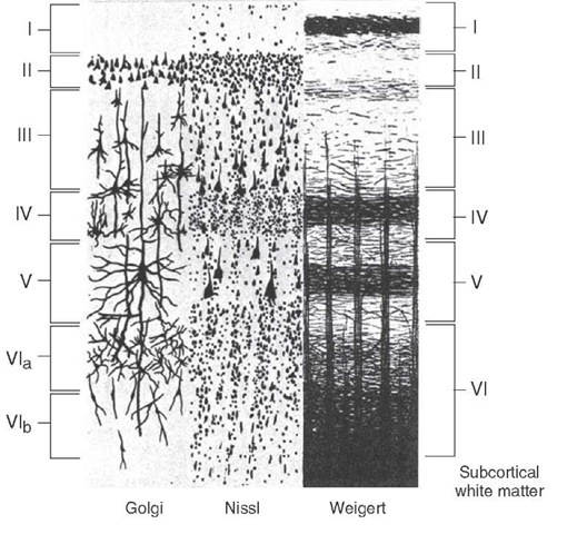
Typically, the cerebral cortex contains six layers arranged parallel to the cortical surface (Fig. 26-1). The layers are generally classified according to the cell type that is most prominent for a given layer (Fig. 26-2). The most superficial layer, layer I, is called the molecular layer. It contains mainly axons, dendrites whose cell bodies are located in deeper layers, and axon terminals of neurons whose cell bodies are also located in deeper layers. The only cell type found in this layer is a horizontal cell, the axon of cells).
FIGURE 26-1 Six-layered structure of the cerebral cortex. The sections are arranged perpendicular to the cortical surface so that the top layer is the one closest to the cortical surface. The illustration on the left depicts the cells as they appear in a Golgi stained section (for identification of neuronal and glial cells and their processes), the illustration in the middle shows the cortical appearance when it is stained for cells, and the illustration on the right depicts the cortex when stained for fibers.
FIGURE 26-2 Intracortical circuits. Note the loci of the synaptic connections (depicted with loops) of afferent fiber projections, the origins of the efferent projections, and the origins of intracortical connections within a given column. The specific region of cortex is not defined in this illustration. G = granule cell; H = horizontal cell; M = Martinotti cell; P = pyramidal cell; S = stellate cell. Arrows indicate direction of information flow.
The most important feature of the granule cell layers (layer II and especially layer IV) is that they are basically receiving areas for inputs from the thalamus and other regions of cortex. Likewise, layers III and V contain mainly pyramidal cells and are thus referred to as pyramidal cell layers. Layer III is called an external pyramidal cell layer, and layer V is called an internal pyramidal cell layer. The pyramidal cell, which is largest in layer V, contains an apical dendrite that extends toward the surface of the cortex and basal dendrites that extend outward from the cell body. The primary efferents of the cortex include axons of pyramidal cells that project throughout the central nervous system (CNS). Layer VI (multiform layer) contains several different types of neurons (spindle-shaped cells with long ascending dendrites, Marti-notti cells with long ascending axons, variations of Golgi cells, and some pyramidal cells [Fig 26-2]). Because of this anatomical arrangement, afferent signals received at almost any layer of cortex can result in excitation of cells located in other cell layers. It is this synchronized form of activation of cells located within a given vertical plane of the cortex that defines the cortical column and thus provides the cortex with its basic functional properties (Fig. 26-2). The concept of a cortical column is discussed again later in this topic in reference to functions of the visual cortex.
FIGURE 26-3 Brodmann’s cytoarchitectonic mapping of the human brain. (A) Lateral view; (B) medial view. The different regions are labeled with corresponding symbols and numbers. Note the key regions that have been identified using Brodmann’s numbering scheme. Several of these regions include: area 4: primary motor cortex; area 6: premotor area; areas 3, 1, and 2: primary somatosensory receiving areas; areas 5 and 7: posterior parietal cortex; area 17: primary visual cortex; and area 41: primary auditory receiving area. See text for further discussion of Brodmann’s areas.
Cytoarchitectonic Division of the Cerebral Cortex
The histological appearance of the cerebral cortex varies from region to region. These variations are commonly associated with functional differences as well. The histological variations were first recognized by Brodmann at the beginning of the 20th century. Consequently, he assigned numbers and symbols to each cortical area to define and characterize the different regions of the cortex (Fig. 26-3). This numbering system has remained in the literature for almost a century and is commonly used to define a given functional region. More recently, specific regions of the brain have been defined in terms of other characteristics, such as their afferent and efferent connections, physiological and receptor characteristics, and relationships to behavioral functions.
As a general rule, histological differences in the appearance of cortical tissue are most apparent when comparing sensory and motor regions of the brain. One of the clearest examples is the difference between the histological appearance of the primary motor (area 4) and primary somatosensory (areas 3, 1, and 2) cortices (Fig 26-3). These differences indicate that, with respect to area 4, the granule cell layers (layers II and IV) are quite small, whereas pyramidal cell layers III and V are rather extensive. In contrast, in a portion of the primary somatosensory cortex (area 3), the granule cell layers are more extensive, and the pyramidal cell layers are reduced in size.
Other examples of cortical regions that contain an abundance of pyramidal cells relative to populations of granule cells include the medial aspect of the prefrontal cortex and anterior cingulate gyrus. The regions that contain an abundance of granule cells relative to pyramidal cell populations include the primary auditory and visual cortices.
Neurotransmitters
Both excitatory and inhibitory neurons are present within the cortex. Excitatory effects may be generated from cortical interneurons that have short projections within a given column or between adjacent columns as well as by thalamocortical and other cortical afferent fibers. Inhibitory interneurons within the cortex are extensive, including those acting within the same column from where they originate or between adjacent columns. Excitatory interneurons are mediated mainly by excitatory amino acids (aspartate and glutamate) that act on glutamate receptors (kainate, quisqualate, and N-methyl-D-aspartate [NMDA]). Other neurotransmitters associated with intrinsic cortical neurons include a variety of neuropep-tides. The neuropeptides include somatostatin, substance P, enkephalin, cholecystokinin, and vasoactive intestinal peptide and presumably serve a modulating function on cortical neurons. With regard to extrinsic neurons, monoamine neurons arising from different regions of the brainstem act on many cell groups within the cerebral cortex, releasing dopamine, norepinephrine, and serotonin from their nerve terminals. These monoaminergic neurons also modulate neuronal excitability levels and play an important role in the processes of sleep and wakefulness and in the regulation of emotional states. In addition, acetylcholine originating from neurons in the basal forebrain supplies wide regions of cerebral cortex. This excitatory transmitter has been linked to functions of learning and memory, and loss of such inputs to the cortex has been reported to occur in disorders such as Alzheimer’s disease.
Cortical Layers Associated With Inputs and Outputs
Different layers of the cortex serve different functions with respect to its afferent and efferent connections. Layer I is primarily a receiving area for nonspecific afferent fibers from the intralaminar thalamus and brainstem monoamin-ergic neurons. Layers II and IV are primarily receiving areas: Layer II is a receiving area for cortical (callosal and association) afferents, and layer IV is a receiving area for afferents from the thalamus. Layers III and V primarily contain efferents: Layer III contains callosal and cortical association fibers, and layer V contains efferents that project to the neostriatum, brainstem, and spinal cord (Fig. 26-4). Concerning layer VI, some of these neurons project to the thalamus, whereas others contain short projections that ramify within the same cortical columns from which they arise.
FIGURE 26-4 Input-output relationships of cortex. Schematic diagram depicts the intrinsic organization and input-output relationships of the cerebral cortex. Excitatory connections are indicated by (+), and inhibitory synapse is indicated by (-). Note that thalamocortical and intracortical projections terminate mainly in layer IV, and monoaminergic projections are distributed mainly to more superficial layers. Cortical afferents terminating in layer IV can either excite or inhibit pyramidal cells in layer V, which contribute significantly to the outputs of the cerebral cortex. The major outputs to the spinal cord, cranial nerve motor nuclei, other brainstem structures, thalamus, and neostriatum arise in layers V-VI, whereas projections to other regions of cortex either on the ipsilateral or contralateral side arise from layer III.
Excitability Characteristics of Neurons Within a Cortical Column
Two factors that regulate cortical excitability should be noted. The first is that large cortical cells, such as pyramidal cells, integrate the activity of many neurons that impinge on these cells. Pyramidal cells also transmit signals to other layers of the cortex by virtue of their axon collaterals. Some connections are excitatory, whereby other neurons within a given cortical column are then excited. Other connections are inhibitory, especially with neurons in an adjacent column. The inhibitory neurons may then make synapse with additional neurons that are either inhibitory (producing cortical disinhibition) or excitatory. Thus, excitation of the pyramidal cell can lead to wide variations in the patterns of cortical excitability. For this reason, the nature of the functional patterns governing excitation within a cortical column still remains poorly understood.
The second factor is that cortical afferents, such as those arising from the thalamus or from other regions of the cortex, have the capacity to activate large numbers of cortical neurons at any one given moment. These sources typically have fast excitatory synaptic actions. Other inputs that are mediated by acetylcholine, monoamines, or peptides appear to have a slow, modulatory-like action on these cells.


