Basal Ganglia
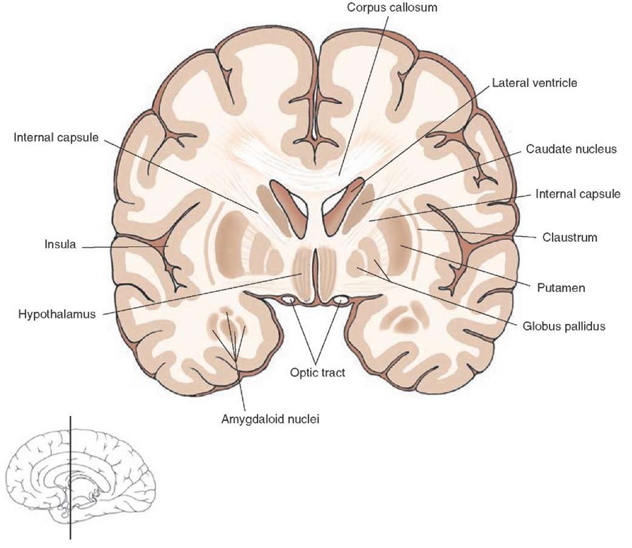
The basal ganglia play an important role in motor integration processes associated with the cerebral cortex. Damage to this region results in motor dysfunctions referred to as dyskinesias (i.e., disorders of movement at rest). The most prominent structures of the basal ganglia are the caudate nucleus, putamen, and globus pallidus (Figs. 1-6 and 1-7). Two additional structures, the subthalamic nucleus and substantia nigra, are also included as part of the basal ganglia because of their anatomical and functional relationships with its other constituent parts.
The caudate nucleus is a large mass of cells that is most prominent at anterior levels of the forebrain adjacent to the anterior horn of the lateral ventricle and can be divided into three components (Fig. 1-8). The largest component, the head of the caudate, is found at anterior levels of the forebrain rostral to the diencephalon. As the nucleus extends caudally, it maintains its position adjacent to the body and inferior horn of the lateral ventricle but becomes progressively narrower at levels farther away from the head of the caudate. This narrow region of the caudate nucleus, distal to the head, is called the tail of the caudate nucleus. The region between the head and tail is referred to as the body of the caudate nucleus. The body and tail of the caudate nucleus are situated adjacent to the dorsolateral surface of the thalamus.
The putamen is the largest component of the basal ganglia and is situated in a lateral position within the anterior half of the forebrain. It is bordered laterally by the external capsule, a thin band of white matter, and medially by the globus pallidus (Figs. 1-6 and 1-7).
The globus pallidus has both a lateral and medial segment. It lies immediately medial to the putamen and just lateral to the internal capsule, which is a massive fiber bundle that transmits information to and from the cerebral cortex to the forebrain, brainstem, and spinal cord (Figs. 1-6, 1-7, and 1-8).
FIGURE 1-7 Frontal section taken through the level of the rostral diencephalon (where the thala-mus is not present). Note again the relationships of the caudate nucleus and diencephalon relative to those of the globus pal-lidus and putamen with respect to the position of the internal capsule. The level along the ros-tro-caudal axis of the brain at which the section was taken is shown in the sketch of the brain at the bottom of the figure.
FIGURE 1-8 Schematic diagram illustrating the components of the caudate nucleus and their relationship to the thalamus, internal capsule, globus pallidus, putamen, and brain-stem. Because of its anatomical proximity to the caudate nucleus, the stria terminalis, which represents a major efferent pathway of the amygdala to the hypothalamus, is included as well.
Diencephalon
As mentioned previously, the diencephalon includes principally the thalamus, situated dorsally, and the hypothalamus, situated ventrally. The medial border of the diencephalon is the third ventricle, and the lateral border is the internal capsule. The ventral border is the base of the brain, and the dorsal border is the roof of the thalamus. The diencephalon is generally considered to be bounded anteriorly by the anterior commissure (Fig. 1-3), which is a conspicuous fiber bundle containing many olfactory and temporal lobe fibers, and the lamina terminalis (not shown in Fig. 1-3), which is the rostral end of the third ventricle. The posterior limit of the diencephalon is the posterior commissure, a fiber bundle that crosses the midline between the diencephalon and midbrain.
Limbic Structures
Limbic structures serve important functions in the regulation of emotional behavior, short-term memory processes, and control of autonomic, other visceral, and hormonal functions usually associated with the hypo-thalamus. Several structures in the limbic system can be identified clearly in forebrain sections. Two of these structures, the amygdala and hippocampus, are situated within the temporal lobe (Fig. 1-6). The amygdala lies just anterior to the hippocampus. Both structures give rise to prominent fiber bundles that initially pass in a posterodorsal direction following the body of the lateral ventricle around the posterior aspect of the thalamus and then run anteriorly, following the inferior horn of the lateral ventricle.
The fiber bundle associated with the hippocampal formation is the fornix, which is situated just inferior to the corpus callosum (Fig. 1-3). The fiber system associated with the amygdala is the stria terminalis and is just ventromedial to the tail of the caudate nucleus (Fig. 1-8). The trajectory of the stria terminalis is parallel to that of the tail of the caudate nucleus. Both fiber bundles ultimately terminate within different regions of the hypothalamus.Other components of the limbic system include the cingulate gyrus, the prefron-tal cortex, and the septal area.
Topography of the Cerebellum and Brainstem
Cerebellum
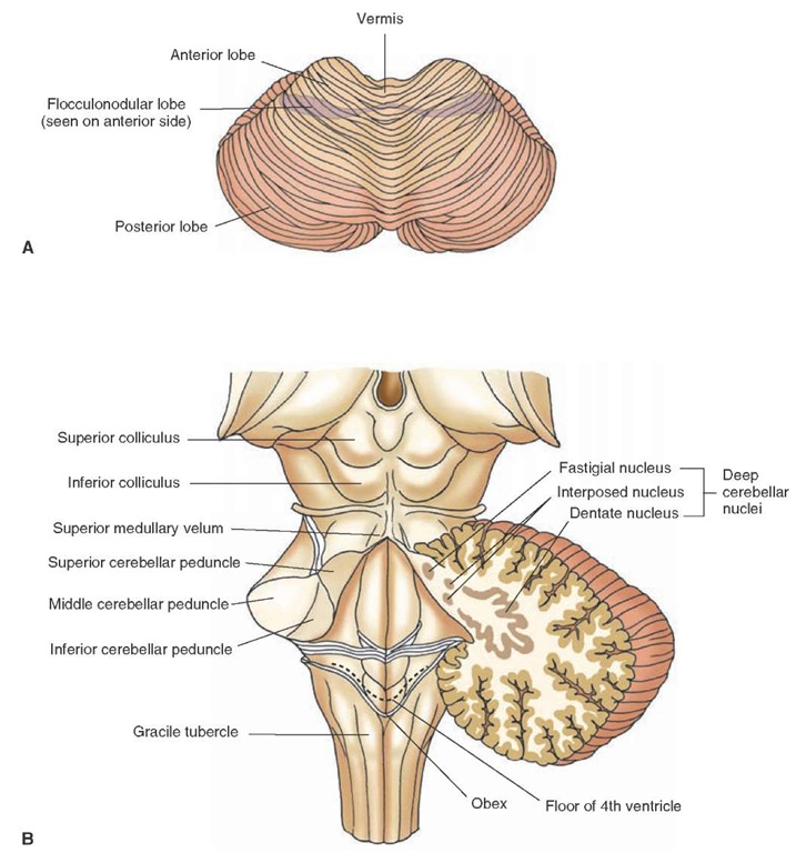
The cerebellum plays a vital role in the integration, regulation, and coordination of motor processes. Damage to this region can result in loss of balance, loss of coordinated movements, hypotonia, and errors in movement when attempting to produce a specific response. It is attached to the brainstem by the cerebellar peduncles, three pairs of massive fiber bundles. One pair, the superior cerebellar peduncle, is attached rostrally to the upper pons. Another pair, the inferior cerebellar peduncle, is attached to the dor-solateral surface of the upper medulla. The third pair, the middle cerebellar peduncle, is attached to the lateral aspect of the pons (Fig. 1-9B).
FIGURE 1-9 Cerebellum and brainstem. (A) Dorsal view of the cerebellum indicating the positions of the anterior, posterior, and flocculonodular lobes and the midline region called the vermis. (B) Dorsal view of the brainstem after removal of the cerebellum. The connections of the cerebellum to the brainstem are indicated by the presence of the inferior, middle, and superior cerebellar peduncles.
The cerebellum contains bilaterally symmetrical hemispheres that are continuous with a mid-line structure, the vermis. The hemispheres are divided into three sections. The anterior lobe is located towards the midbrain. Extending posterior-inferiorly from the anterior lobe is the posterior lobe, the largest lobe of the cerebellum. The flocculonodular lobe, the smallest of the three lobes, is situated most inferiorly and is somewhat concealed by the posterior lobe. It is important to note that each of these lobes receives different kinds of inputs from the periphery and specific regions of the CNS. For example, the floccu-lonodular lobe primarily receives vestibular inputs, the anterior lobe receives inputs mainly from the spinal cord, and the posterior lobe is a major recipient of cortical inputs.
Brainstem
Dorsal View of the Brainstem
Two pairs of protuberances at the level of the midbrain can be seen on the dorsal surface of the brainstem (Fig. 1-9B). The superior colliculus is more rostrally positioned and is associated with visual functions; the more caudally positioned inferior colliculus is associated with auditory processing. The dorsal surface of the pons and medulla form the floor of the fourth ventricle (Fig. 1-9B). The walls of the ventricle are formed by the superior cerebellar peduncle, and the roof of the fourth ventricle is formed by the superior medullary velum, which is attached to the superior cerebellar peduncle on each side.
In the caudal half of the medulla is the end of the fourth ventricle, the position at which the ventricle becomes progressively narrower and ultimately continuous with the central canal that continues into and throughout the spinal cord. The position at which the fourth ventricle empties into the central canal is the obex. The part of the medulla that contains the fourth ventricle is the open medulla, and the part that contains the central canal is the closed medulla. On the dorsal surface of the caudal medulla are two protuberances, the gracile and cuneate tubercles (cuneate tubercle is situated immediately lateral to gracile tubercle but is not labeled in Figure 1-9). These contain relay and integrating neurons associated with ascending sensory fibers from the periphery to the medulla.
Ventral View of the Brainstem
Crus Cerebri. A massive fiber bundle passes from the cerebral hemispheres into lower regions of the brainstem and spinal cord at the level of the midbrain (Fig. 1-10). This fiber bundle is the crus cerebri, part of the descending complex of motor pathways that communicates signals from the cerebral cortex to the brainstem and spinal cord.
FIGURE 1-10 Ventral view of the brainstem. Note the positions of the cerebral peduncle; basilar part of the pons; pyramid; pyramidal decussation (situated immediately rostral to the cervical spinal cord); inferior olivary nucleus, which lies lateral to the pyramid; and root fibers of cranial nerves.
Pons and Medulla. The pons has two parts: a dorsal half, the tegmentum, and a ventral half, the basilar pons. The basilar region forms the anterior bulge of the pons seen on a ventral view of the gross brain (Fig. 1-10). The tegmentum can only be seen on horizontal or cross sections of the brainstem.Several protuberances separated by a narrow fissure can be detected at the level of the medulla. Starting from the medial aspect, one protuberance, which passes in a rostral-caudal direction along the base of the brain, is called the pyramid. The axons contained within the pyramid originate in the cerebral cortex and thus represent a continuation of many of the same fiber bundles that contribute to the internal capsule and parts of the cerebral peduncle at the levels of the cerebral hemispheres and midbrain, respectively. In this manner, the pyramid serves as the conduit by which cortical signals pass to all levels of the spinal cord for the regulation of motor functions.
The pyramid can be followed caudally from the pons through most of the medulla. At the caudal end of the medulla, near its juncture with the spinal cord, the pyramid can no longer be easily seen from a ventral view. This is because most of the fibers contained within the pyramid pass in a dorsolateral course from the lower medulla to the contralateral side through a commissure, the pyramidal decussation. This pathway, referred to as the lateral corticospinal tract, descends to all levels of spinal cord.The pyramidal (motor) decussation is more clearly visible from a cross-sectional view of the caudal medulla. A small sulcus separates the pyramid from a more lateral protuberance, the olive. The olive is formed by the inferior olivary nucleus, a large nuclear mass present in the rostral half of the medulla (Fig. 1-10). The olive represents an important relay nucleus of the spinal cord and regions of the brainstem to the cerebellum.
Other important features of the ventral surface of the brainstem are the roots of the cranial nerves.These cranial nerves include the oculomotor (cranial nerve [CN] III) and trochlear (CN IV) nerves at the level of the midbrain; trigeminal (CN V), abducens (CN VI), and facial (CN VII) nerves at the level of the pons; and nerves VIII, IX, X, and XII at the level of the medulla (not labeled in Fig. 1-10).


