Introduction
When human remains are rendered unrecognizable by advanced decomposition or are completely skeletonized, experts must be able to make the four determinations:age, sex, race and stature. This information is the most essential aspect of the identification process and indeed, gives police a starting point for their investigation. Of these demographic characteristics, the estimation of age at death is the most challenging once adulthood had been reached. As in the living or recently deceased, precise estimation of age without documentation such as a birth certificate or driver’s license is not a simple task because of the tremendous variability in the manifestations of the aging process. How humans look as they age is orchestrated by the complicated interplay of genetic, environmental, and cultural factors. Everyone’s hair does not turn gray at the same age or rate. The same goes for the development of wrinkles and other features associated with advancing age. Moreover, a face lift and hair dye can make someone look much younger, whereas smoking and sunbathing will add years. Even though plastic surgery does not alter the appearance of the sites that osteologists depend on for estimating age at death, the skeleton also shows the effects of age, and these are just as subject to variation.
There are, however, some basic biological processes that all humans undergo from the prenatal months through infancy and childhood and finally to adulthood. If one understands the growth process, it becomes possible to separate early childhood from 1 to 12 years and adolescence from 13 to 18 years. Adulthood can also be divided into young, middle and older stages. The events of these periods are also reflected in the skeleton. For example, early childhood is characterized by dental growth and development, adolescence by completion of bone growth, middle age by remodeling and maintenance that gradually decline to the wear and tear of old age.
The aim of this article is to present some of the basic dental and skeletal changes that can be used as age markers in an examination of unknown remains. The literature on age estimation can be found in several topics and leading review articles.
Dental and Skeletal Growth
Bone growth and dental development have been studied by skeletal biologists interested in the skeletal remains of children in both prehistoric and modern populations. During the early years, there are basically two major physiological activities in progress: dental development and eruption, and bone mineralization and ossification, both of which hold important clues to the age of an immature person. Under normal circumstances, the order of events during the developmental stages follow regular, predictable patterns with little variability.
The most reliable way to estimate age in children is based on the sequence of both deciduous and permanent tooth formation and eruption. These are excellent age markers because there is consistency in the timing and sequence of eruption. Of the dental sets, the deciduous dentition is first visible at about 6-7 months and is completed by 3 years (Table 1). During this time, eruption begins with the central incisors in the mandible and ends with the second molar. Although the sequence is well established and rarely varies, population variation exists in the timing of dental eruption and this has been studied in many different parts of the world. Tables 1 and 2 display ages of eruption for deciduous and adult dentition, respectively, in several populations. Some researchers claim that accuracy can be best estimated from the enamel formation in the deciduous dentition (up to age 4 years) and in the ensuing permanent teeth as well as the root.
Table 1 Age of eruption (in months) for deciduous teeth
| Population | 11 | 12 | ml | m2 | |
| Maxilla | |||||
| Egypt | 9.4 | 11.2 | 19.1 | 14.1 | 24.7 |
| Tunisia | 9.3 | 12.5 | 18.3 | 15.7 | 22.0 |
| USA | 7.5 | 8.0 | 18.0 | 14.0 | 25.0 |
| Canada | 9.0 | 10.2 | 18.0 | 15.2 | 27.5 |
| New Guinea | 10.6 | 11.9 | 18.2 | 16.0 | 24.1 |
| Mandible | |||||
| Egypt | 8.3 | 12.2 | 19.1 | 14.1 | 24.7 |
| Tunisia | 6.0 | 14.0 | 19.0 | 15.7 | 21.7 |
| USA | 6.5 | 7.0 | 18.0 | 14.0 | 25.0 |
| Canada | 7.1 | 12.0 | 18.2 | 15.3 | 26.5 |
| New Guinea | 8.2 | 13.0 | 18.8 | 16.6 | 23.9 |
Figure 1 contains a sketch of the development of both deciduous and permanent teeth. It indicates the level of tooth development and time of emergence at a given age. As can be seen in this figure, Ml is the first permanent tooth to erupt. Observations of the roots reveal that the canine and M2 are the last of the regular sequence to be completed. In general M3 is not used in age estimation because of the enormous variation in the timing of its eruption. One must also bear in mind that there is a continuing microevolutionary reduction in the dental apparatus a major feature of which is the trend toward impaction and congenital absence of ‘wisdom teeth’, especially in Whites. It should be noted that Fig. l gives general age ranges for the stages of development rather than precise times for these events and does not account for sex and race differences. All research on this topic has clearly demonstrated that the sequence of development and eruption begins earlier in females and Blacks. There is no question that the dental sequence is the most reliable and accurate means of age assessment during the first l2 years of life.
Table 2 Age of eruption (in years) of permanent dentition
| Country | Upper | Lower | Upper M2 |
|||
| I1 | C | P1 | P2 | |||
| Boys | ||||||
| Czech | 7.2 | 8.1 | 11.2 | 10.4 | 11.0 | 12.1 |
| Egypt | 8.0 | 9.1 | 12.1 | 11.3 | 12.1 | 12.7 |
| England | 8.1 | 8.9 | 12.4 | 11.4 | 12.1 | 12.4 |
| Hungary | 7.3 | 8.5 | 11.4 | 11.8 | 11.9 | 12.4 |
| USA | 7.5 | 8.7 | 11.7 | 10.8 | 11.4 | 12.7 |
| Japan | 8.0 | 9.2 | 11.6 | 10.8 | 11.6 | 12.9 |
| Girls | ||||||
| Czech | 6.9 | 7.7 | 10.6 | 9.4 | 10.6 | 11.7 |
| Egypt | 7.3 | 8.6 | 11.8 | 10.8 | 11.2 | 11.7 |
| England | 7.7 | 8.7 | 11.0 | 11.4 | 12.0 | 12.1 |
| Hungary | 7.1 | 8.0 | 11.6 | 10.0 | 11.3 | 12.0 |
| USA | 7.2 | 8.2 | 11.0 | 10.2 | 10.9 | 12.3 |
Bone formation and growth can also be used as an age indicator in the immature skeleton. The formation of bone tissue (ossification) proceeds in a systematic and organized manner. Beginning at loci referred to as centers of ossification, development normally follows a well-known sequence and timing of events culminating in the development of the diaphysis and epiphysis. Yet, as is the case with most human biological functions, there can be variability in all aspects of this process, primarily in timing, and to a lesser extent, in sequence. During adolescence, the cartilaginous growing region or metaphysis of the endochondral bones is replaced by bone to unite the epiphyses with the diaphyses.
Much of our current knowledge of bone formation and development has been derived from radiological studies, and age-related skeletal changes have been studied from the fetal stage through late adolescence. Prenatally, age estimation is based on size differences in each of the many fetal bones. Growth proceeds at its greatest velocity during intrauterine life and infancy, but slows considerably once the 2nd year is attained. The next major growth spurt occurs at puberty accompanied by the development of secondary sex characteristics.
Fontanelle closure in the infant skull is another source of clues to age at death. The posterior fontanelle begins ossifying at 2-3 months after birth and closes by about 6 months, whereas the anterior fontanelle shrinks after birth but does not close completely until about 2 years of age. The metopic suture which runs from nasion to bregma on the frontal bone normally closes by around the age of 6 years. In rare instances, it remains open throughout life.
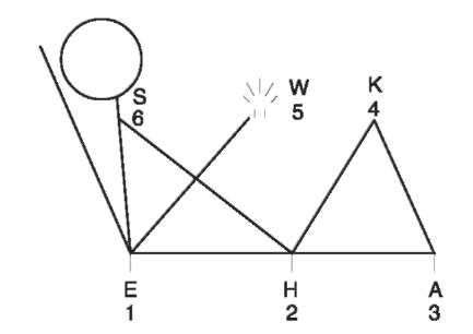
As with the dentition, bone growth is also subject to variability by sex and population. Radiological growth charts are available for several populations to assess age of epiphyseal growth and union. However, there are very few direct studies of skeletal remains because of the scarcity of children in documented collections. On average, epiphyseal fusion begins at about 12-14 years. This process starts earlier in females as a component of their earlier attainment of sexual maturity. The first joint of the body to unite is the elbow formed by the articulations of the distal humerus and proximal radius and ulna. Fusion then proceeds in a set order throughout the skeleton until growth is complete sometime between the late teens and early 20s with males active at the older ages. Figure 2 is a simplified sketch showing the order of epiphyseal fusion beginning first in the elbow and ending in the shoulder. In general, there is about a one year differential between each of the joints; thus it normally takes about 6 years to complete epiphyseal union ranging from about 12-14 years to approximately 18-20 years. Again, sex and population differences must be taken into account. By the early 20s, most of the epiphyses are united with the exception of the basisphenoid synchondrosis of the skull which has been observed to close as late as 29 years and the medial clavicle which may remain open into the 30s. Although a long bone length-based age estimation has been suggested, growth in adolescence is so much more varied than that characterizing the intrauterine months and early childhood, that epiphyseal union is by far the most reliable age marker in the teens. The best estimates are obtained if figures have been drawn from the same population as that of the skeleton in question.

Figure 1 Ages of development and eruption of deciduous teeth (A) and permanent dentition (B). (Modified from Massler ef a/. (1941) Developmental pattern of the child as reflected in the calcification pattern of teeth. American Journal of Diseases of Children 62: 33.)
Adult Age
Unlike the regular predictable order of growth and development in childhood, once maturity is attained, skeletal structures are modified and maintained by the process of remodeling. This activity can be highly variable from one individual to the next and the alterations produced are usually subtle and often difficult to detect and interpret. The adult aging process is susceptible to the influences of human behavior and the environment. This is clearly seen in the fact that even genetically identical twins can differ from each other as they get older. Thus, age estimation in the adult is one of the most challenging assessments to make from the skeleton, and middle age is probably the most difficult because it is an extremely varied transitional period hormonally and metabolically. The later years also have their share of complications, including inevitably higher rates of pathologic conditions and the effects of a lifetime of wear and tear. Of particular concern to the forensic anthropologist is the fact that few skeletal components show discernible patterns of age-related change past the 40s.

Figure 2 Order of epiphyseal closure beginning with the elbow joint (E) at about 12-14 years of age, followed (at approximately 1 year intervals) by the hip (H), ankle (A), knee (K), wrist (W), and ending with the shoulder (S) by 18-22 years.
The first attempts at the development of adult age estimation techniques from systematic studies of (supposedly) known aged skeletons began in the 1920s with observations of the pubic symphysis and cranial sutures. These were followed by years of attempts to improve accuracy at these sites. Unhappily, cranial suture closure is so inherently erratic that it remains a technique of last resort and only used when no other alternative is possible. For many years and with many modifications, the pubic symphysis had been considered more reliable, but now experts have conceded that it has a limited range of usefulness. It is problematic by the 30s and ineffective by the 40s. Nearly every part of the skeleton has been examined for adult age assessment, but few sites have proven to be consistently reliable. Different methodological approaches such as radiography and histology have also been attempted, but with limited success.
It took over 60 years until serious attention was shifted to any other part of the skeleton than the cranium and pubic symphysis. Today the most consistently reliable method for the determination of age at death from the adult skeleton is based on a lifetime of changes at the sternal end of the rib that have been divided into a series of phases beginning in the late teens. Standards for white males and females have been extensively tested with consistently accurate results on many diverse modern and archaeologic populations. Table 3 shows that accuracy ranges from as little as +1.5 years through the 20s to about +5 years thereafter. The youngest phase (0) and oldest (8) are open ended (e.g. less than 17 or over 70).
The aging process in the rib was assessed by studying changes at the costochondral junction of the fourth rib (Fig. 3). The metamorphosis of the rib begins after the completion of growth at the sternal extremity at about 14 years in white females and 17 in their male counterparts. This is consistent with the generally earlier maturation of females. It is characterized by the disappearance of the epiphyseal line and the beginning of pit formation at the nearly flat, billowy surface of the sternal end of the rib. Within a few years, the pit has taken a definite V-shape and scallops appear on the rim of the bone. The pit gradually deepens and widens to a wide V (in females) or U shape in both sexes. The rounded edges of youth begin to thin and sharpen by the mid 30s. With increasing age, the rim becomes irregular, the interior of the pit becomes porous and the bone quality deteriorates until the ribs of most individuals over 70 years are thin and irregular with bony projection at the costochondral junction.
Table 3 Comparison of mean ages and 95% intervals for rib and pubic symphysis phases
| Phase | Rib | Pubic symphysis | ||
| Mean | 95% Interval | Mean | 95% Interval | |
| Males | ||||
| 1 | 17.3 | 16.5-18.0 | 18.5 | 15-23 |
| 2 | 21.9 | 20.8-23.1 | 23.4 | 19-34 |
| 3 | 25.9 | 24.1-27.7 | 28.7 | 21-46 |
| 4 | 28.2 | 25.7-30.6 | 35.2 | 23-57 |
| 5 | 38.8 | 34.4-42.3 | 45.6 | 27-66 |
| 6 | 50.0 | 44.3-55.7 | 61.2 | 34-86a |
| 7 | 59.2 | 54.3-64.1 | ||
| 8 | 71.5 | 65.0-a | ||
| Females | ||||
| 1 | 14.0 | 15-24 | ||
| 2 | 17.4 | 15.5-19.3 | 25.0 | 19-40 |
| 3 | 22.6 | 20.5-24.7 | 30.7 | 21-53 |
| 4 | 27.7 | 24.4-31.0 | 38.2 | 26-70 |
| 5 | 40.0 | 33.7-46.3 | 48.1 | 25-83 |
| 6 | 50.7 | 43.3-58.1 | 60.0 | 42-87a |
| 7 | 65.2 | 59.2-71.2 | ||
| 8 | 76.4 | 70.4-a | ||
There are considerable race and sex differences in bone density with age. Bone density decreases dramatically in Whites, and especially females, whereas it remains minimally changed in Blacks, with the least deterioration in males. However, the same process that contributes to the maintenance of density over age 30 (periosteal deposition) also makes the rib look older because it creates irregular bony projections in Blacks (particularly males) decades before these features appear in Whites. Concerns over side and intercostal differences have been pretty much dispelled. Several studies have shown that although standards are based on the right fourth rib, there are no side differences and intercostal variation among true ribs 2-7 and especially between ribs 3, 4 and 5 is not significant.
Figure 3 Rib cage phases 0-8: metamorphosis of the sternal end of the rib in males (M) and females (F). The solid, smooth immature rib has a flat or billowy end with rounded edges bordered by the epiphyseal line (phase 0). This site undergoes a series of changes characterized by the formation and deepening of a pit at the costochondral junction, accompanied by thinning and porosity of the bone and development of sharp, irregular edges throughout the life span to Phase 8 in extreme old age (over 70 years) (see Table 4). (Modified from iscan and Loth (1989) The osteological manifestations of age in the adult. In iscan MY and Kennedy KAR (eds) Reconstructions of Life from the Skeleton, pp. 23-40. New York: Wiley-Liss.

One reason the rib phases are quite precise and reliable is the fact that sex and race differences were investigated and accounted for in the standards. Since, as noted earlier, these factors produce significant variation in the rate and pattern of rib metamorphosis through the lifespan. Moreover, there is little individual variation because the only direct stress on the ribs is breathing. This contrasts with other skeletal sites like those in the pelvis which are directly involved with weight bearing, pregnancy and parturition.
Although the rib is the bone of choice, the sternal end is not always present or intact so the forensic anthropologist must be prepared to evaluate whatever information is available from other skeletal components. In addition, the odds of correct age estimation increase if other sites yield similar results.
Before the development of the rib phase method, the pubic symphysis was considered the most reliable morphological technique. The general aging pattern at this site is represented in Fig. 4. Standards have been reworked numerous times to attempt to cope with great variability, and casts have been created to help improve the effectiveness of matching the sym-physis to an age phase and reduce interobserver error. However, the results of independent tests of several pubic symphyseal techniques were found to be disappointing in accuracy and precision. Furthermore, as can be seen in Table 3, even though a bone can be matched more easily to a cast, the 95% confidence intervals of the age range is too large to be very useful for most phases.
The latest pelvic site to be tapped is the auricular surface of the ilium. Developed for use on archaeologic samples where it was possible to seriate a large number of specimens, the workers in this area have reported good results on early cemetery and American Indian burials. However, this method is very difficult to apply and independent testing indicated that this site was not suitable for use in individual forensic cases.


Figure 4 Schematic of pubic symphyseal changes with age.
Often only a skull is found or made available to the forensic anthropologist for analysis. Unfortunately, there are no precise age indicators in this part of the skeleton and one can only give a very rough estimate such as young adult, middle aged or elderly. In the skull, the cranial suture closure pattern and its relation to age has been rigorously investigated for nearly a century. In spite of the considerable attention paid to this technique, variation is so enormous that it is practically abandoned as an age marker. It is not all that rare to see an entirely fused skull suture in the 20s or patently open ones in the elderly. It has been suggested that because a suture is a long structure it can exhibit a range of intrasutural variability along its length. In an attempt to reduce this problem, the most recent method offered a modified approach. Instead of examining the entire suture, only 1 cm sections were selected for assessment at specified locations. The closure pattern at each spot was scored as follows:
Score 0: Completely open. No ectocranial closure. Score 1: Minimal to moderate closure, from a single bony bridge to about 50% synostosis. Score 2: Significant, but not complete closure. Score 3: Complete obliteration and fusion.
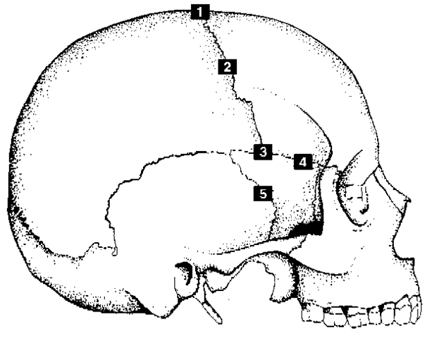
The five lateral-anterior suture sites (1-5 in Fig. 5) yielded the best results and the total score can be converted into an age estimate using Table 4. The mean ages using this technique ranged from 32 to 56 years. It must be emphasized, however, that the developers of these standards note that there is still great variability and recommend that this site be used in conjunction with other indicators. Attempts have been made to correlate age with the closure of other sutures of the vault and facial skeleton, but results have not been satisfactory.
The association between tooth wear and aging has long been known and is still commonly used for archaeologic assemblages. This process has been studied by many scientists for nearly 100 years. It is a generally reliable indicator in relatively small, ancient populations that were genetically and nutritionally homogeneous for whom group specific rates of attrition could be calibrated from juvenile samples.

Figure 5 Suture closure at lateral-anterior sites (taken at points indicated by numbers). 1. Bregma; 2, midcoronal (midpoint of each half of coronal suture at pars complicata); 3, pterion; 4, sphenofrontal (midpoint); 5, sup. sphenotemporal 2 cm below juncture with parietal bone.
Table 4 Age estimates from lateral-anterior suture composite scores
| Composite | Mean | Interdecile | ||
| Score | N | Age | SD | Range |
| 0 (open) | 42 | |||
| 1 | 18 | 32.0 | 8.3 | 21-42 |
| 2 | 18 | 36.2 | 6.2 | 29-44 |
| 3-5 | 56 | 41.1 | 10.0 | 28-52 |
| 6 | 17 | 43.4 | 10.7 | 30-54 |
| 7-8 | 31 | 45.5 | 8.9 | 35-57 |
| 9-10 | 29 | 51.9 | 12.5 | 39-69 |
| 11-14 | 24 | 56.2 | 8.5 | 49-65 |
| 15 (closed) | 1 |
The great individual variation of contemporary humans reduces the effectiveness of this site for aging. However, one of the most recent examples of efforts to use dental attrition for clues to age today appears in Fig. 6. Although the standards were developed from archaeologic Amerindians, application to turn of the twentieth century American Whites and Blacks produced surprisingly good results. The figure depicts discernible stages from A through I (in both jaws), that begin as early as 12 years and continue as late as 55 years and reports no sex differences. Cross-hatching signifies the transitional stage from the enamel to the secondary dentine formation. The black areas indicate that the enamel is entirely gone and dentine is visible. It is clear that the wear pattern goes from the anterior to the posterior teeth.
Tooth wear is likely to have the best relationship to broad or general age categories of any other indicator in the skull, but its application to forensic cases must be interpreted cautiously because (as noted above) of the tremendous individual variation in current large, heterogeneous, modern populations. These differences encompass many factors including relative enamel thicknesses, significant racial dimorphism, widely varied diet, socioeconomic status, and access to health and dental care. A further consideration is that, although the people of today live considerably longer than their archaeologic counterparts, wear rarely progresses to the extreme stages seen in the figure. This is primarily due to our relatively soft refined diet, free from the grit that literally ‘sanded away’ the entire tooth crown. In addition, tooth loss, resulting from decay and periodontal disease, is much more common now, and more so in Whites than Blacks.
Another, more comprehensive approach to dental aging in the adult was first introduced nearly 50 years ago. It was designed to assess a number of characteristics of a cross-section of a tooth including attrition, periodontosis, secondary dentine, cementum apposition and root resorption. Since the original study there have been many modifications of this technique. Although the method can yield a reasonable age range estimate, its disadvantages include the removal and destruction of a tooth (usually a canine or premolar), and the laboratory equipment and time necessary for thin sectioning and microscopic analysis.

Figure 6 Age phases of dental wear in the maxilla and mandible (see Table 4). (From Lovejoy (1985), Dental wear in the Libben population: its functional pattern and role in the determination of adult skeletal age at death.
Other parts of the adult skeleton can sometimes be used to assign an upper or lower value. In the absence of trauma or disease, the development of osteophyticlipping on the bodies of the vertebrae is a good indication that an individual is over the age of 40 years. Bone quality can also be a guide. Firm solid bones are consistent with youth whereas thin, porous, fragile bones signal old age.
Radiologic and Histomorphometric Techniques
Radiology is not commonly used by anthropologists and few real standards are available. Some of the earlier studies go back to the 1950s when epiphyseal involution was observed. Currently, the proximal epiphysis of the humerus, femur and tibia are all well documented. Again as in many other age estimation techniques the deterioration of the spongy tissue has been sequenced and divided into several stages. Several other bones such as the clavicle and ossification of costal cartilages have also been observed. Radiography may be effective at giving a general idea of age from a mature long bone where there is no other noninvasive alternative, but it cannot be relied upon for precise estimates.
Age estimation using histomorphometry was first experimented with in the 1960s. This technique attempts to correlate age with the cellular activity involved with bone remodeling at the histologic level by counting osteons, assorting them into different stages of development or breakdown, and calculating their ratios. The remodeling process has been studied by many investigators who found it to vary by race and sex. Population specific standards have been introduced for both contemporary and prehistoric populations. The age range covers the adult stage of human life span. Accuracy, as measured by the standard error of estimate claimed in some publications on the subject varies by around 6 years. Quantified comparisons of rib phase assessment with histomorphometric analysis from the same sample demonstrated that sternal end phase morphology was at least twice as accurate.
Microscopic techniques have a number of disadvantages. Technically, this is a very laborious and time-consuming procedure requiring specialized training. Osteon counts are subject to a great deal of interobserver error. Even among experts, assessment of the osteons and related structures varies considerably and is thus subject to high levels of interobserver inconsistency. This technique also requires destruction of the long bone shaft or rib which must be cut to prepare thin sections. Therefore, this approach must be pursued with caution when dealing with forensic cases. It is not routinely used and is usually limited to specialists.
Discussion
Estimating age at death continues to be one of the most challenging aspects of skeletal analysis because of the complexity and individual variation of the aging process and the myriad of factors that affect it. Osteologists use the term ‘estimate’ because we can get close to chronological age – as with the rib phases which provide a meaningful range in which there is a high probability (95%) that true age will fall. It cannot be overemphasized that age estimates must be expressed as a range rather that erroneously using the mean value as a point estimate. To do so can lead to the elimination of the actual victim from being considered as a match for unknown remains.
Although scientists have gained a good idea about the general stages of age changes in the skeletal system that hold true for the most part, there are numerous factors that accelerate or decelerate the process. This can result in unexpected morphologic manifestations that may cause errors in age estimation. Human variation, on both the individual and population level is significant. So race and sex differences must always be considered in age estimation standards and decisions.
When estimating age from the skeleton, one must remember that what has been analyzed in research is the skeletal age rather than birth certificate-based chronological age. Researchers attempt to reconcile the internally regulated developmental and remodeling processes in the skeleton with the passage of time. Conflicts (errors in age estimation) arise because the former follows its own ‘biological clock’, and the latter is fixed by the regular, unalterable ticking of the atomic clock. To make matters worse, growth, development and remodeling can proceed at different rates in different parts of the skeleton. In other words, one component may not clearly reflect the sum total of skeletal development. Chronological age represents time based or true age and attempts to elucidate this depend on expected bone formations at a given point in time. However, a person at a particular level of skeletal development may have reached that stage earlier or later than the average. The physiologic processes that underlie aging are dependent on many internal and external influences including genetic make up, health and nutritional status, and substance abuse. During growth the situation becomes especially complex when age estimation is linked to bone lengths in children because no matter how much variation is incorporated, the potential is there to make errors. The same applies to remodeling in adulthood. This is why it is so important to provide age ranges so that extremes are not omitted from the police list of missing victims.
Finally, it must also be kept in mind that some skeletal sites and methods are more accurate than others. However, forensic personnel must be cognizant of all possible options since one rarely has a complete skeleton or even the bone of choice.
