Introduction
Dust is the microscopic, particulate residue of a universe that is crumbling under the forces of nature and human beings and has been described by the great chemist Liebig as matter in the wrong place. Because microscopic particles are characteristic of their source, easily transferred between objects and persons, and as transfer is difficult to prevent, dust has special significance in forensic science. It was through study of and experimentation on dusts of all types that Dr Edmond Locard of Lyon laid the foundation for the collection, analysis and interpretation of contact traces in the first half of the twentieth century. The results of this work were formulated into what L C Nickolls called Locard’s exchange principle: ‘Whenever two objects come into contact there is always a transference of material.’ This principle is the basis and justification for the detection, recovery and analysis of microscopic trace evidence. Since the quantity of matter which may have been transferred is frequently quite small, and since all transferred material is subject to loss from the surface to which it was transferred, over time and through motion, these tasks must be performed by a trace evidence analyst who is equipped both by training and experience to perform these tasks in a thorough, scientific and efficient manner.
Goals and Uses of Dust Analysis
As one might expect, both the goals and techniques of dust analysis have changed since Locard’s time. Locard’s experiments were directed primarily toward identifying the occupation and geographic movements of a suspect from an analysis of the dust from clothing, earwax, fingernails, etc., and using this information to help solve crimes. In one case, for example, he isolated the dust from the clothing of suspected counterfeiters. Microchemical analysis showed that the metallic particles comprising the majority of the dust consisted of tin, antimony and lead, the very metals of which the counterfeit coins were composed. The suspects’ normal occupations would not have produced particles such as those that were found on their clothing. Today’s trace evidence analyst uses microscopic evidence to establish links in various combinations between a victim, suspect and crime scene. For example, fibers similar to those from the clothing of a suspect may be found on the body of a victim, or soil similar to that at the scene of the crime may be recovered from a suspect’s shoes. Less often, the laboratory is asked to provide investigative leads to detectives in the field. In these cases, the scientist may be asked, for example, to determine the likely origin of particles found on the bodies of the victims of a serial killer. This was the case with colored paint spheres recovered from the naked bodies of murdered prostitutes in London between 1964 and 1965. These particles led the police to a repaint shop to which, as it turned out, the killer had access. In another example, the origin of sand, substituted for a rail shipment of silver flake between Colorado and New York, was determined, on the basis of a mineralogical examination of the sand grains, to have originated from a location near Denver.
One can see from the examples above that the analysis of microscopic particles has the potential to provide a wealth of useful information during the investigation of a crime and the trial that follows after a suspect has been identified and charged. The principal questions that can be addressed by the analysis of dust fall naturally into three main categories, although others may suggest themselves in certain circumstances.
1. Is there evidence of likely contact, in some combination, between a victim, suspect and the scene of the crime} This is the most commonly asked question and accounts for most courtroom testimony involving microscopic trace evidence. If Locard’s exchange principle holds true, there must be a transfer of material if contact was made. It is important to remember, however, that the inability to detect physical evidence of a contact does not mean that no contact occurred. As someone once wisely said, ‘Absence of evidence does not mean evidence of absence.’ Even through a transfer of material occurs, the quantity may be so small as to escape detection, or the decay rate may be such that evidence is lost before it is brought to the laboratory for examination. Another possibility related to quantity is mixing. This is a particular problem with certain types of trace evidence, such as soil taken from the wheel well of a vehicle, where mixing of various soils may make it impossible to reach a conclusion regarding the identity of a source. Fibers are the type of particulate evidence most commonly collected and studied. This is due in great part to three factors. The first is the common occurrence of fibers in our modern environment from clothing, carpets and upholstery. The second is the publicity surrounding a number of high profile murder investigations that were solved and prosecuted primarily with the aid of fiber transfers, such as those associated with the Atlanta child murders in the United States. The final reason is that confirmation of Locard’s theory was experimentally proven in the last 30 years of the twentieth century by using fibers as transfer objects. In fact, many other types of particles may be transferred in a contact; thus the forensic microscopist who studies dust must have developed the ability to identify a wide range of particles by their microscopic characteristics. As an example, the discovery and analysis of minute, green spheres of spray paint, which had been sprayed on cotton fibers to color the headliner of a car, were the most significant piece of physical evidence linking the suspect to the crime scene in the oldest murder retrial in US history.
2. Can the origin of a dust sample be discovered, or its location described, from its composition} The answer to this question is often of practical significance in an investigation. Whether or not it can be answered depends on both the nature of the dust involved and the skill and experience of the scientist conducting the analysis. Dust, by its very nature, always contains clues to its origin. These clues may not always be found, their significance may be obscure or they may not have importance to the case at hand, but certain generalizations may be made in almost every case. Thus indoor dusts normally contain large numbers of fibers. If the source is a home, these fibers will be of textile origin and include fibers from clothing, carpets, curtains, upholstery, etc. Human hairs, as well as animal hairs from pets and feathers from bedding or pet birds, are also commonly found. The composition of the dust will also be controlled by the particular room from which the dust originates. Thus, the bath and bedroom will contribute large numbers of skin cells, pubic hairs and particles originating from talcum powder, antiperspirants, cosmetics, soap, etc. Dust from the kitchen contains particles of spices, flour, starch, etc.
Homes are not the only type of indoor location. The dust from an office may contain many of the same particles found in the living areas of a home. However, the fibrous component of an office dust tends to be dominated by the presence of paper fibers rather than those from textiles. Dusts of outdoor origin, on the other hand, consist primarily of silt-sized mineral grains, pollen, spores, plant cells and tissue, leaf hairs, soot and charred particles from plant burning. The exact composition of an outdoor dust depends on its geographic source, whether it is urban or rural, the time of year it was collected and the types of industries at or near the source. It is possible to narrow the range of inquiry by specifically identifying components of the dust and then studying their peculiarities. For example, it is possible to determine if quartz sand originated from a dune in a desert or from a seashore, on the basis of the shape and surface texture of the individual quartz grains comprising the sand. The identification of the mineral grains as quartz would be made using the petro-graphic microscope; then the surface of these grains would be examined with the scanning electron microscope. If the sample were large enough to isolate some of the trace (accessory or heavy) minerals, it would be possible to draw further conclusions regarding its geological origin once they had been identified with the petrographic microscope. Thus, although beach sands from the east and west coasts of the United States look similar at first glance, their accessory minerals are different: those from the west originating from the relatively young Rocky Mountains and coast ranges; and those from the east, especially the southeast, originating from the old metamorphic rocks of the Appalachians. If our sample also contained pollen grains which, after identification, would give us an idea of the flora in the region, it should be possible to narrow the possible sources of the sand to a relatively small geographic region. Additional particles present in the dust may make it possible to describe the source more closely still. While this example shows the possible lengths to which such an examination can be taken, it is important to realize that the true origin may be complicated beyond what a sound scientific analysis can provide. Thus, the most recent source of this hypothetical sand might be from a pile at a construction site and not the beach where it was formed. Fortunately, sands used for civil engineering projects are not usually transported over great distances and the conclusions would still generally be correct. Still, this illustrates the pitfalls to which conclusions drawn from this type of dust analysis are subject and points out the need for caution in the interpretation of the results.
3. Is it possible to determine a person’s occupation from an examination of the dust on clothing’: This was one of the principal concerns of Locard’s original studies and he reported great success in this area. While it is undoubtedly true that people do pick up dusts from their place of work according to the exchange principle, the nature of these dusts may not be particularly distinctive and, when they are, they may be deposited on a set of clothes or uniform which the worker only wears when on the job. Thus, office workers carry paper fibers on their clothing, but so do people working in other offices, and the style and quality of the clothing itself may indicate this without the need to analyze the dust. Occupations that produce or expose workers to distinctive particulate matter still exist, and in these cases characteristic particles may be carried to vehicles and back to homes if the subjects do not change and wash at their workplace. In a recent specific example of this, the home of the murder victim contained significant numbers of metal turnings, which were brought into the home by her son, a machinist, who stopped off frequently to visit after work.
Collection and Recovery of Dust Traces
The collection of dust traces at a crime scene must be carried out with great care, as the exchange principle applies equally to criminals, victims and evidence technicians. The most effective trace evidence laboratories either collect their own evidence or train specialists to collect evidence in an efficient and scientific manner, while at the same time taking precautions to prevent contamination as the evidence is being collected and packaged. These precautions extend to wearing a white cleanroom garment, hat and booties over the clothing when collecting evidence from the crime scene. When multiple sites or vehicles are being investigated, the evidence collector must change to a fresh set of barrier clothing to prevent possible contamination from occurring between crime scenes. Failure to take such precautions has caused legitimate concern about contamination to be raised in cases where positive trace evidence comparisons were made because evidence was carelessly collected. For example, in a bank robbery where two vehicles were used in succession during the getaway, evidence was collected by investigators who visited and collected evidence from both of the vans in the same day while wearing the same normal street clothing. It is impossible to state categorically that contamination could not have occurred under these circumstances.
It is possible, in some cases, to seek out suspected traces of dust from specific locations or from items of clothing or vehicles by direct examination. In other instances it is necessary to collect the dust first and then sort through it later to search for fibers or particles that may have a bearing on the case. In a recent homicide, for example, the murderer removed iron grillwork from a brick wall to enter the victim’s home through the window. Microscopical examination of the suspect’s clothing revealed particles of apparent red brick dust. Subsequent analysis of these particles proved that they were brick and that they were, both in color and petrography, the same as the bricks from the wall from which the grill had been removed. In this case the facts that the suspect’s clothing contained brick dust and that his occupation did not involve working with brick illustrated the truth of Louis Pasteur’s statement that ‘chance favors the prepared mind’.
There are several methods of collecting dust for analysis. Perhaps the simplest and most direct is hand-picking, with the aid of a stereomicroscope or loupe, using forceps or needles. This was the way in which the suspected brick particles were removed from the clothing in the previous example. The only other necessary aids are a source of bright light and steady hands. Hairs, fibers or particles recovered in this manner should be immediately placed on glass slides, in well slides or small Petri dishes, depending on the size of the item, and covered. The location and type of particle as well as the date and the location from which it was removed should all be noted. If at all practical, the specimen should be photographed in situ before it is isolated for analysis.
Other methods of collection are available for recovering dusts of smaller particle size than those that can be easily manipulated with forceps. Selection of one of these methods over another will depend on the item or location from which the particles are to be recovered and the purpose for which the dust is being collected. If, for example, the substrate is an item of clothing and it is being searched for evidence of recent contact, taping the surface with transparent adhesive tape is the method of choice. Experiments have shown that this is the most efficient method for recovering surficial dusts. This technique was developed by a student of Locard, the late Dr Max Frei-Sulzer of Zurich. It has the advantage that the tapes can be placed sticky-side down on clean slides or acetate sheets, thus preventing contamination while at the same time providing a clear view of the collected dust when searching with either a stereomicro-scope or automated fiber finder. The sticky tape method is also of use when a specific but relatively small area is to be searched for trace evidence. Thus, the area around a point of entry into a building can be sampled efficiently by pressing tape around a window frame, broken glass, torn screen, etc. to collect any traces from the clothing of the person who passed through the opening. Occupational and environmental dusts which have penetrated the weave of clothing over time, and large areas such as the interior of a vehicle, may require the use of a vacuum cleaner. Other dust collection methods such as washing and scraping may occasionally be of value. Washing is useful when the particles are in a sticky or greasy matrix. Locard described a case in which he washed the hair of suspected bomb makers in a gallon of ethanol. When the alcohol evaporated, it left a thick residue of nitrocellulose, the principal component of smokeless powder. Scraping is rarely, if ever, used in European laboratories but is still in use in some laboratories in the United States. The item is hung over a large sheet of fresh paper and a large metal spatula is scraped over the surface. The dust that falls down on to the paper is collected and placed in small containers for later sorting under a stereomicroscope.
Analysis of Dust Traces
The complete analysis of a sample of dust can be a time-consuming and difficult exercise in micro-analysis, but fortunately a full dust analysis is rarely required for forensic purposes. It is necessary at the outset of the analysis, however, to formulate clear and concise questions that the analysis is expected to answer. Thus, if the question were ‘Are there any blue-dyed rabbit hairs from the victim’s sweater on the suspect’s clothing or in his car?’, it would be necessary to first characterize the fibers from the sweater so that similar fibers might be recognized in the tapings from the suspect’s clothing and vehicle. Such fibers are called target fibers and they give the microscopist something to look for in trying to answer the question.
Identification of the components of a dust presents special challenges because of the small size of the individual particles. While some substances can be recognized at sight under the relatively low magnification of a stereomicroscope, most microscopic particles must be isolated and mounted between a slide and coverslip in a specially chosen mounting medium. Particle manipulation is performed under a stereomi-croscope using finely sharpened forceps and specially sharpened tungsten or steel needles, which have points much finer than those used in common dissecting needles. Once mounted in a familiar mounting medium, many particles can be recognized, at least to their class, by a well-trained and experienced forensic microscopist. The ability to identify a large number of particles on sight or by using a few simple optical tests is an important skill and useful advantage for the trace evidence analyst. It means that considerable time can be saved and the microscopist can quickly focus in on the particles that may be most helpful in answering the questions that have been posed. Even the most experienced and skilled worker will still require the aid of other techniques and instruments to confirm an identity and, if necessary, perform comparisons of particles from dust. Some of the most useful techniques and instruments for the analysis of the components are listed and described briefly below.
Microscopy
Many particles can best be identified on the basis of certain morphological characteristics. Substances of this type usually belong to a group in which all the members are chemically similar. An example of this is cellulose. Wood and wood fibers, the nonwoody vegetable fibers, rayon and lyocell fibers all have nearly the same chemical composition, yet it is a simple matter to distinguish each of these groups from one another and within a group to identify the individual members on the basis of their microscopical characteristics. Botanical fragments from leaves, seeds, bark, flowers, stems and roots are all identifiable, with suitable training and reference materials, and in the same way are pollen grains. No amount of chemical analysis would identify these substances as accurately as a well-performed microscopic analysis. As for comparison, it is a general rule that the best comparison of such materials is an accurate identification.
The optical properties of amorphous or crystalline transparent particles and manmade fibers, measured or estimated with the aid of the polarizing microscope, extend the range of application of the microscope to the identification of materials without regard to their morphology. Thus glass, minerals, drugs and chemical crystals of all types can be identified, when encountered as single small particles, on the basis of their optical properties. The technique, when applied by a skilled microscopist, has the same advantages as X-ray diffraction, in that it is possible to identify a particle as being a particular phase and not just a particular compound. One could distinguish, for example, between gypsum (calcium sulfate dihy-drate), anhydrite (calcium sulfate) and plaster of
Paris (calcium sulfate hemihydrate) on the basis of the optical properties measured on a single small crystal. Aclassical chemical analysis of each of these compounds would show only calcium and sulfate ions, while X-ray spectroscopy would show only calcium and sulfur. Polarized light microscopy is particularly useful for rapidly identifying manmade fibers as a prelude to comparison.
Microchemical analysis
It is frequently necessary to obtain chemical information from small particles to confirm their identity, or to identify metallic particles or unusual organic or inorganic compounds that cannot be identified with certainty by microscopy. Classical microchemistry is a useful skill, as microchemical testing can be performed on inorganic, organic or metals with equal ease and speed on very small particles. Although microchemical testing with reagents has been largely supplanted by instrumental methods, such tests, when applied by a scientist trained in their use and application, are rapid and provide chemical information that may be difficult or impossible to obtain with other techniques. The detection of acid radicals or organic anions can be performed with certainty, the oxidation state of an element can be identified, and tests are available for classes of compounds (e.g. carbohydrates), to name just a few of the many possibilities. The principal types of microchemical reactions are based on crystal tests, spot tests and histochemical reactions that have been scaled down so that they can be applied to single small particles or small quantities of material. Crystal tests are usually performed on a microscope slide. The reaction products are crystalline compounds with characteristic crystal form, habit or optical properties. Spot-test reactions result in reaction products that are highly colored, gaseous or fluorescent. Positive histochem-ical tests usually result in the formation of highly colored or strongly fluorescent reaction products which form in situ on the particle itself. The value of microchemical testing in dust analysis is that it can rapidly provide chemical information on extremely small particles with very little cost in money or time.
X-ray spectroscopy
This technique is capable of providing an elemental analysis of extremely small particles and is thus well suited to the analysis of dust particles. The spectrometer is attached to a scanning electron microscope. The electron beam of the instrument generates X-rays from the specimen in addition to the imaging secondary and backscattered electrons. The energy of the X-rays are related to the atomic numbers of the elements in the sample. In modern instruments, all of the elements, from boron up, are detectable. The advantage of this technique is that the analysis is simultaneous and the entire elemental composition of an extremely small particle (down to about 0.1%) can be obtained in seconds. It makes no difference if the sample is inorganic or organic, so both metals and paints, for example, can be analyzed with equal advantage.
Infrared microspectrophotometry
Just as the elemental composition of a small particle can be obtained from an X-ray spectrometer, its molecular composition can be determined by this instrument. It consists of a Fourier transform infrared spectrophotometer attached to a reflecting microscope. An infrared spectrum permits the analyst to identify organic compounds and anions by comparison to reference spectra or by interpretation of the infrared absorption bands in the spectrum. The microspectrophotometer makes it possible to collect the spectrum from particles as small as a few micrometers in diameter. Although there are many other sensitive instruments in the modern analytical laboratory, these have not, as yet, been employed to any great extent in dust analysis.
Reference Collections
Reference collections are essential for the forensic science laboratory that conducts dust analyses. These collections are composed of physical specimens of the small particles which comprise the world in miniature. The number of specimens and completeness in any category will vary by laboratory. In some areas the collections are built over time; in others they will have been acquired by purchase. The items to be included will depend on the availability and the interests of the analysts in the laboratory. Potential items for inclusion are too numerous to list completely, but a useful collection might include the following: metals (both different metals and alloys as well as metal particles which have drilled, sawed, crushed with a sledgehammer, etc.), industrial dusts, combustion products, paper (woody) and nonwoody vegetable fibers, manmade fibers, human and animal hairs, minerals, sands (from different geological environments), wood, pollen, spores, diatoms, leaves, seeds, food products (e.g. spices, grains, starches, spray-dried products), polymers, synthetic drugs, explosives, dusts from different environments (these can be collected from air filters in hotel rooms or by vacuuming particles from the clothing after returning from a trip), and occupational dusts (obtained by vacuuming the clothing of volunteers working in various jobs involving manual labor). Depending on the nature and the amount of sample available, items may be stored in small vials, envelopes or as permanent mounts on microscope slides. An entire collection may thus be stored in a several drawers in a few relatively small cabinets.
EAR PRINTS
Historic Overview
It is only during the last few years that ears and ear prints, and the identification of (unknown) ear prints found at a crime scene and known ear prints from a suspect, have attracted the attention of the police and prosecuting attorneys. Nevertheless, the use of ears and ear prints as a means of establishing identity is quite old.
In olden days, for instance, the length of the ear lobe was considered to be a sign of great wisdom in some countries in Indo-China, which is why statues of Bhuddha always have long lobes. Aristotle considered long ear lobes to be a sign of a good memory. During the Renaissance, when physiognomy was introduced and the followers of this doctrine pointed out that the face is a reflection of all intelligent qualities of a human being, much attention was paid to the shape of the ear. During his research into primates, Darwin also attracted the attention of the scientific world by saying that the ear is one of the elementary organs. In support of this, he pointed to the broadening of the middle of the helix, indicating that this is nothing else but a corner of the primitive ear that has become reduced in size. Science acknowledged this by naming it the ‘tubercle of Darwin’.
Schwalbe was one of the first to invent a method of measuring the external ear. He was able to prove Darwin’s theory and was the first to attract scientific attention to racial peculiarities in the structure of the ear.
Quotes from others who expressed their feelings about the importance of the ear can be found in Die Bedeutung der Ohrmuschel fur die Feststellung der Identitdt (The Meaning of the Auricle for Identification Purposes) by Imhofer of Prague. He quotes Lavater (1741-1801), who, in publications during the period 1775-1778, argues for the position of the ear to be considered as part of physiognomy. Imhofer also quotes Amide Joux, saying: ‘Show me your ear and I’ll tell you who you are, where you come from and where you’re going.’ The author refers to his conviction that there is no other organ of the human body that can better demonstrate the relation between a father and his child. It was his belief that the shape of the ear is clear proof of paternity. The article continues by stating that ears can be very important in establishing identity in:
• the determination of the identity of corpses;
• the determination of the identity of living persons; for example, when establishing the identity of a wanted person, or establishing family relationships.
Imhofer quotes Bertillon by writing that: ‘It is almost impossible to find two ears which are equal in all parts and which have many shapes and characteristics which stay without noticeable changes throughout a life time.’ The article continues with a discussion of methods for comparing ears and the characteristic features necessary to determine identity, as well as the numbers of characteristic aural features found in comparative research, with 500 pictures of ears. A combination of three features only appeared in two cases and it was concluded that a combination of four features is enough to establish identity.
In the development of a system to protect parents, children and hospital administrations from cases of mistaken identity in babies, Fields and colleagues suggested using the appearance of the ears in newborn babies. Footprints, palmprints and fingerprints, in conjunction with some additional identification, were generally used in most hospitals. The problem with these prints was that there was no or little reference value for identification and the prints were often of such poor quality as to render them useless, even within the first month. After a test study, taking pictures of infants, it was noticed that the appearance of the ear remained constant, whereas other features of the face changed. When a questionnaire was sent to plastic surgeons throughout the English-speaking world, 75% of the responses favored the use of the ear as a constant for identification.
A study was conducted and included photographs of the right and left ears of a series of infants, taken daily from the day of birth to the day of discharge from hospital. These photographs were taken to document the minute changes that took place in the growing ear during each 24 h period and to document the gross morphologic changes that took place from the date of birth to the date of departure from the hospital. The result of the research was that 206 sets of ears were photographed and the following conclusions were reached:
• The ears of different sample babies are always unique; no baby was found to have ears identical in size or configuration to those of another.
• There is enough variation in individual ear form to distinguish visually the ears of one baby from those of another.
• The sample series of the photographed ears of a baby remained homogeneous (markedly similar in form and structure) during the entire hospitalization period.
• The minute changes that take place in the growing ear are so slight that the immutability of the series is not jeopardized.
The described photographic research procedure was deemed to have met all of the requirements of a reliable and standardized identification technique: individuality, continuity and immutability.
The identification of individuals by ear characteristics has been described in a text for scene-of-crime officers by Soderman and O’Connell. The principal parts of the ears are described, as well as peculiarities of the helix and the antitragus, descriptions that were said to be useful for identification purposes. An ear print case was presented by Medlin, giving a description of the way the case was handled and the results. The actual procedure of the comparison is not explained.
The characteristics of the auricle, measurements of the ear and morphological characteristics with genetic importance have been studied by Oliver, who described variations in the form of the ear, the darwinian tubercle and lobular adherence.
Oepen presented the morphological aspects of ears, in a lecture and articles for the German Forensic Medicine Society, from research on the differences between the features of the ears and their appearance in practice in 500 men and women. The study was done to find out what features of children’s ears were characteristic and could indicate the identity of the biological father. The outcome of the research was a useful contribution to the comparison of ears. Several tables illustrate the findings regarding aural features and give statistical information about how often a combination of features was found. Other tables show the features that were found more often in women then in men, and vice versa, and more often on the right side than on the left side, and vice versa. Attention is paid to the way in which an ear will print and what features of the ear will be visible.
Hirschi from Bern in Switzerland dealt with a burglary case in which ear prints were found. The ear prints of a left and right ear could be identified as placed by suspect ‘P’. Besides the information as to on what ground the prints were identified, the report also contains quotes from the well-known and respected scientists Reiss, Niceforo and Locard regarding the value of ears in the identification process. Hirschi also gives reference prints from test persons that were used for the comparison. He describes the work that had been done regarding the height at which the ear print was found and the height of the perpetrator. Hirschi carried out research among 40 recruits from the Swiss Police Academy as to the degree to which they bent over; the distance between the top of the skull and the middle of the auditory canal; and whether they were right-handed or left-handed.
Hunger and Leopold discussed medical and anthropological views regarding the identification of people and referred to the shape of the ear being basically specified by its cartilage. They pointed out that the external ear has many specific features useful for identification, particularly because these features are very stable throughout the aging process and even after death. They note that not all of the characteristic features may be recognized on photographs, which also applies to prints.
Trube-Becker from Dusseldorf pointed out that there are no absolutely identical ears, but only similar ears. Even two ears of one and the same individual are not completely identical. This is equally true for identical twins. A warning is given with respect to the fact that the properties of ears can change as they are pressed against a wall or a door. Nevertheless, as they are clearly recognizable, comparison is possible. The opinion is stated that ear prints can at least provide additional circumstantial evidence for a suspect’s guilt and that it is therefore worthwhile looking for ear prints and preserving them like fingerprints. They can provide an additional piece in the mosaic of identification of the perpetrator.
Georg and Lange described the case of a bank robbery in which a comparison was made of several features of the human body for identification purposes. They expressed the view that multiple morphologic variations (not only in facial characteristics but also differences in, for example, the features of the hands and fingernails, height and body shape) are at least of the same value in identification as fingerprinting. The ear in particular provides many morphologic and metric features: as with fingerprints, no two people in the world will have ears that have the same combination of features. On the basis of several morphologic features in this particular case, a suspect was apprehended. Statistical calculations showed that the combination of morphologic features in this case could only be found 1 in 300 000 000 people.
Cases involving ear prints in the surroundings of Dusseldorf have been investigated by Trube-Becker. She renders an opinion on several crime scene prints and states that many of these prints originated from a suspected perpetrator ‘with a probability bordering on certainty’. She describes the work that was done by the Ludwigshafen Criminal Investigative Police, who apparently found ear prints repeatedly. The prints were rendered visible with soot dust or a Magna Bush and lifted with foil. For making the standards (controls) from suspects they developed a system using a Polaroid CU-5 camera with a transparent macrolon disk on its front. They were able to place the disk against the head and actually see the change in the form of the ear caused by the pressure. The prints were then made visible with fingerprint powder and lifted with foil. At the same time the ear was photographed at a scale of 1:1. A built-in ring flash allowed a colored picture to be taken but it appears that the flash is always visible on the photograph, which has such a damaging effect on the result that the photograph cannot be used for the purpose for which it was intended.
There have been cases in Freiburg (Germany) with findings of ear prints in a series of burglaries. Meyer relates a case in which two girls were seen near the site of the burglaries. Police suspected them of having listened at the door before breaking in. The girls were arrested for identification purposes, during which prints of the left ear were taken. The next day a message was received from Stuttgart, where three girls had been arrested for the same type of burglary. Prints of these girls’ left ears were also taken. Investigations quickly showed that (according to the ear prints) the girls from Stuttgart could not be excluded from having committed the burglaries in Freiburg. The prints from the scenes of crime were taken to a fingerprint expert in Baden-Wiirttemberg, who stated that: ‘The traces found at the different crime scenes were identical and caused by a left ear. The prints contained more or less the same part of the ear corresponding to the cartilage of the ear. The traces of the ear prints contained each at least 8 of the 12 most important points of the ear that can be notified.’ A comparison was made between the prints from the crime scenes and the standards from the girls. It was found that the prints were identical to the prints from one of the girls arrested in Stuttgart. The expert stated: ‘Between the standard of the left ear and the three prints from the crime scene there is, taking into account a tolerance defect, the highest match in form of coverage equality’.
The anatomical design of the auricle is so individual and multiple that, with a probability bordering on certainty, it can be stated that no two ears can be found that are identical in all details. Hammer summarizes the value of ear print identification by saying that, of 100 ears investigated, no two ears could be found to correspond in all features and that the human ear print is therefore suitable for establishing a person’s identity in forensic practice, although the reliability of the information it gives on questions of identity depends on the quality of the ear prints secured at the scene of the crime.
Hunger and Hammer, from many years’ practical experience, stated that the ear can play an important role in the identification of both perpetrators and unidentified corpses. They describe the results of research into the distribution of selected ear characteristics in the East German population, recording which types of aural features were found to be rare in a group of 350 men and 300 women. The authors also made two concluding observations. First, as regards the identification of the ear from a severely dismembered corpse, special care must be taken in preparing the ear for preservation by photography. It appears that characteristics can be altered when the tissues in the head area are separated from their surroundings. The second point concerns ear prints, of which it is said that when the ear is pressed firmly against a surface, the ear can change, particularly in size and shape. Hunger and Hammer state that the metric and morphologic characteristics of the ear are very well suited for determining identity, especially when rare characteristics are present.
The first case in which ear prints led to a conviction by a Dutch court of law, reported by Dubois, concerned a burglary and hostage-taking. The scene of crime investigation and the procedure leading to the ear identification are described, together with the examination of 100 test subjects to find out whether or not their ears were similar. This research showed that the ear of none of the test persons resembled the ear found at the scene. In fact, great dissimilarities were found, whereas the ears of a suspect were identical. The brothers of the suspect were asked to contribute to the investigation to find out whether the suspect’s ears were similar to his brothers’ ears. The results were very much to the contrary. During the investigation a forensic odontologist and an ear, nose and throat specialist were consulted. The court at Dordrecht accepted the ear prints as evidence and convicted the suspect. The court of appeal in The Hague only used the ear print as supporting evidence and convicted the suspect, partly on the basis of this but mostly on other evidence in the case.
Rochaix has said: ‘The ears are after the fingerprints the best means for identifying people, because they stay the same from birth till death. They do not change except for the size. The ear is the most characteristic feature of the human being’. He also describes the form and features of the human ear and points to the work done by Bertillon, Reiss and Locard, as well as the method used by the cantonal police at Vaud (Switzerland) for recovering and lifting prints and taking the standards from a suspect. He introduces a classification method for ear prints based upon several features of the ears, which will usually be visible after listening. In total there are five features to be used for obtaining a final classification code; these items include the shape of the ear; the attachment of the ear lobe to the cheek; the bending and basis of the antitragus. These codes give a series of numbers for both left and right ears and can therefore be stored and found easily. Rochaix concludes that, using this classification, 600 different ear prints can be stored separately. Whether or not this system is adequate to separate the different ear prints found needs verification.
Hammer and Neubert summarized their research and that of Hirschi with respect to measurements of height. They found that the distance between the top of the skull and the middle of the auditory canal was 13.1 cm for men and 12.4 cm for women in the East German population. Similar values were found in a study in 1986, namely 13.2 cm for men and 12.5 cm for women. They conclude that it is only possible to ascertain the height approximately because: (1) the middle of the external ear cannot always be determined; (2) the inclination of the head while listening averages 3 cm (according to Hirschi) but the range is 1-8 cm; (3) although average ear height might be known, there is about 4 cm between the minimal and maximal values. The authors also give the results of research into pressure distortion. The differences between ears that were pressed weakly and strongly against a surface were investigated at eight different points of the ear and differences in four descriptive ear characteristics were noted. Among the 100 ears that were analyzed, not one of them was completely identical to another.
Kritscher and colleagues described the identification of a bank robber partly based upon comparison of the ear. The available information came from a surveillance camera, which showed a man of medium height with only the right ear clearly discernible and thus useful for identification purposes. They describe the method and measurements used for comparison. The images of the bank robber’s right ear, as caught by the surveillance camera, and the suspect’s right ear proved identical in virtually all respects. The authors concluded by saying that their morphologic-anthro-pometric examinations enabled them to ascertain the identity of the bank robber to a large degree, in particular as the matching morphology of the auricles could be rated as strong a proof as are identical fingerprints.
There are those who doubt the possibility of identifying a person by their ears or ear prints. Moenssens gives his basic reason for doubt by stating that there has been no empirical research on the subject. On the other hand he writes: ‘A close look at the external part of the ear shows that it contains numerous complicated details which may be arrayed in particular combinations possibly unique to each person.’ He continues by saying: ‘Forensic anthropologists recognize the possible individuality of an individual’s ear, but not as a means of identification via ear impressions.’ Attention is given to the fact that there is a significant difference between comparing actual ears and photographs of ears, and comparing ear prints to each other; and it is concluded that a reliable and replicable standard procedure has yet to be developed. There have not been any professional conferences on the subject, nor any court decisions at the appellate level recognizing such a discipline and admitting evidence based upon its comparisons.
In Poland a study of 31 cases involving ear prints was carried out over a 5 year period. Labaj and Goscicki give advice on how to recover and lift ear prints and how to take into account the height at which the print is found. They use a distance of 17 cm to arrive at the estimated height of the perpetrator.
A literature survey and practical research on the influence of pressure on ear prints has been carried out at the National Training Centre at Durham, UK. Saddler has reported on ear prints, with an overview of the materials and the methods used to obtain them, an explanation of the method used for measuring and a discussion on the development of a computerized database.
The present author’s own research on the relation between the height of an ear print at the scene of a crime and the height of the perpetrator was similar to the research done by Hirschi in Switzerland. It was carried out between 1990 and 1992 at the Dutch College for Criminal Investigation and Crime Control at Zutphen. The difference between the two investigations related to the number of test subjects used in the research and the fact that the Dutch research studied both men and women and used a different method. The findings of this research were also different. The distance between the top of the skull and the middle of the auditory canal appeared to be 14 cm on average for men and women; the inclination was on average 6 cm.
In Rumania, ear prints are also used for identification. Pasescu and Tanislav give an overview of a number of cases handled since 1975. The number of cases increased after the publication in 1976, by the Institute for Forensic Studies, of a practical handbook of forensic investigation, which included a synthesis of the main scientific observations on ear prints that had been published up to that time. They describe the sites at which ear prints were found and the way in which they were recovered and lifted.
Although ear print evidence has been used in practice in the Netherlands since 1986 (the Dordrecht case), and more frequently since 1992, the attention paid to this type of evidence (although it has been included in scene-of-crime officer training since 1991) increased after a murder case in 1996 in England, followed by one in Washington State. The American case included a FRYE-hearing about the admissibility of ear prints as well as the trial itself. The case led to the first admission of ear print evidence in the United States, as well as the first conviction based upon this evidence.
Morphology of the Ear
Basically the ear consists of cartilage, which gives the original shape and dimensions to the ear, covered with skin. The development of the ear starts shortly after conception and by the 38th day some of the features of the ear are recognizable. The ear moves to its definitive position on about the 56th day and the shape of the ear can be recognized on the 70th day. The shape is normally fixed from then on and never changes from birth until death.
Figure 1 illustrates the external features of the ear. Ears can be divided into four basic shapes: oval, round, rectangular and triangular (Fig. 2). These shapes occur in all races but the percentage of each shape differs between races. The difference in the shape of ears established from ear prints is just a class characteristic.
For comparative purposes, the individual appearance of the features of the ear, their dimensions and their relation to other features are of most importance. A combination of features that can be identified can lead to the individualization of the ear print.
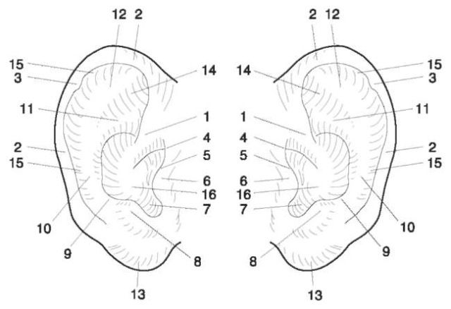
Figure 1 The external features of the ear: 1, crus of helix; 2, helix; 3, auricular tubercle (of Darwin); 4, anterior notch; 5, anterior tubercle; 6, tragus; 7, intertragic notch; 8, antitragus; 9, posterior auricular furrow; 10, anthelix; 11, lower crus of anthe-lix; 12, upper crus of anthelix; 13, lobule; 14 triangular fossa; 15, scaphoid fossa; 16, concha.
Where to Find Ear Prints
Experience shows that ear prints are found predominantly on surfaces where an individual has been listening to determine whether or not a premises was occupied. This occurs generally at doors or windows;hence the surfaces that are likely to be examined are predominantly glass, wood or painted (Fig. 3). However, it may not be at the door or window where entry was gained, or even the same premises, that an ear print is found: individuals may have listened at several premises along a street, or several windows in one house.
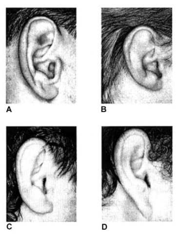
Figure 2 Ear shapes: (A) oval; (B) round; (C) rectangular; (D) triangular.
In the Netherlands the majority of ear prints are found on doors in blocks of flats. They are found at two different heights: one close to the ground and one at normal standing height. Within any block of flats, ear prints may be found on doors and windows on one floor or several floors, not all premises having been entered.
The first procedure, which is always utilized when attempting to recover ear prints from any type of surface, is a thorough visual examination. It is essential that there is good lighting, either natural or artificial, and considerable care is necessary to carry out a thorough and effective examination. Artificial lights should ideally produce a high level of flat and even illumination, rather than the glare produced from some sources. Additionally, combinations of change, involving the angle of light, the viewing position and where possible the rotation of the article, will significantly affect the success rate. Some ear prints can only be seen clearly by the use of oblique light. Care is needed when handling articles using this technique so as not to damage other ear prints that may not be visible at this stage.
The latent impressions can be made visible by applying a suitable reagent, e.g. fingerprint powder.
In the case of ear prints that are fresh, the powder principally adheres to the aqueous component of the sweat deposit, whereas with older ear prints fatty deposits become much more relevant.
Traditionally, the most widely used and productive method of recovering fingerprints is the application of fingerprint powder, followed by photography or lifting of the marks found. This method can also be readily applied to ear prints (Fig. 4).
Influence of Pressure
Because of the influence of varying pressure, ear prints recovered from a crime scene will never be identical in every respect to standards (or controls) taken from a suspect. This is due to the fact that the exact pressure exerted at the crime scene is an unknown quantity and therefore cannot be reproduced. Likewise the direction from which the pressure was applied cannot be duplicated exactly. Although it is impossible to recreate the exact pressure of the ear and rotation that left the impression, it is possible to create a similar impression so that pressure features and other characteristics may be compared. In fact a ‘duplication’ of the amount of pressure is not necessary.
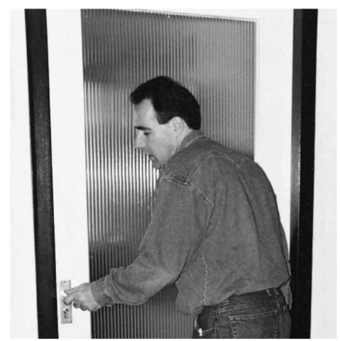
Figure 3 The manner in which someone listens at a door.
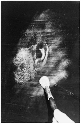
Figure 4 Application of fingerprint powder to an ear print.
In summary, unlike fingerprints, where identical marks are left, no ear print impression will ever be identical but it will have points of similarity that can be compared.
Depending on the applied pressure, features of the human ear may be visible, or not yet visible. By applying more pressure it is likely that more features of the ear become visible, depending on the individual characteristic features of the ear. Features that were already visible will become clearer when more pressure is applied. Through exertion of pressure the so-called ‘pressure points’ of the ear will become visible and may leave a characteristic mark. To be able to study the changes in the appearance of the ear by application of pressure, at least three different standards or control samples need to be taken from a suspect. These must illustrate distinct pressure variations. By studying the differences caused by pressure, one can see or predict how an individual ear will change with pressure.
In addition to pressure, the effect of rotation of the ear when listening at a door or window must be taken into account.
Producing Standards (Controls) from Suspects
Several different methods have been employed to produce standards from suspects in an attempt to identify perpetrators. Most documented methods for obtaining standards include photographing the ear, in addition to any other technique employed. If a suspect cooperates in the investigation, standards should be made as follows.
The suspect is asked to ‘listen’ three times at a glass pane in a door (Fig. 5). The pressure that is exerted on the pane must be different each time: the first print taken is from the application of gentle pressure; with normal pressure; and the third with much more pressure (Fig. 6).
This method of taking prints is used for both right and left ears. In all cases a photograph of both ears is taken, with the camera at an angle of 90° to the head (Fig. 7).
The prints thus obtained are made visible in the same way as are fingerprint identification traces, and lifted with black lifter.
For suspects who will not cooperate, and those who offer passive resistance, standards are taken using glass or a synthetic plate (Fig. 8). Five prints of each ear must be made, putting variable pressure on the ear while preventing one part of the ear (upper, lower, front or back) having extra pressure placed upon it.

Figure 5 Taking standards (controls) from a cooperative subject.
Procedures for Comparison
The procedure for comparison has two aims: first, to establish whether or not the ear print in question contains enough features for a comparison; and, second, to be able to tell the court that a strict procedure was followed every time an ear print was compared. This procedure consists of six steps.
Step 1 (analysis)
Note down every detail of the print in question:
(a) What type of fingerprint powder (or chemical) was used to reveal the latent print?
(b) What type of lifter was used to lift the print?
(c) What features of the ear are visible? (Use a picture with anatomic features to note down the details.)
(d) Which of the features might be specific for these ears?
(e) What pressure points are visible on the earprint?
(f) What additional information is visible? For instance: skin texture of cheek, head, etc.; hair; scars, pimples or birthmarks.
Is the information enough for comparison with a known print?
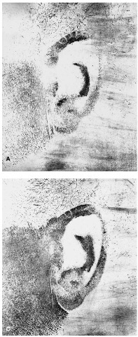
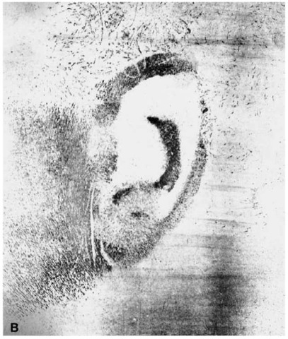
Figure 6 Prints left by (A) light pressure; (B) medium pressure; (C) heavy pressure.
Step 2 (analysis)
The same process applies to the known prints (the standards taken from a suspect). Follow step 1 (a)-(f); in addition:
(g) Is there enough difference in pressure between the three (or five) ear prints to be able to say something about the way pressure influences these ears?
(h) What are those differences?
(i) What parts become visible when more pressure is applied?
(ii) What parts become more dominant when more pressure is applied?
(iii) What about changes in the overall form and dimensions?
Step 3 (preparation)
Depending on the type of lifter, a number of steps must be performed to obtain a result that can be photocopied.
• Black gelatine foil When the ear print has been lifted using a black gelatine foil, the color must be inverted: further comparison requires a gray/black ear print image on a white surface. A ‘dustflash’ is an apparatus that enables transfer of the image directly on to a photographic paper on a 1:1 basis. Photographic development will reveal the image.
• White gelatine foil This type of foil is not recommended for ear print lifting. The powder that is usually employed is a black powder (a soot powder) that does not adhere very well to the latent print. If this type of foil is used, the procedure to follow depends on the visibility of the print. Good prints can be photocopied directly; in other cases the print should be photographed and printed (on a 1:1 basis).
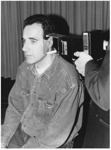
Figure 7 Photography of the ear.
• Transparent foil or tape This type of foil (either used in combination with a gray aluminum powder or a black soot powder) can either be directly photocopied of photographed.
In all cases a digital image can be made, providing the print is reproduced on a 1:1 basis.
Step 4 (preparation)
Photocopies of both the known and unknown ear prints are produced on plain and transparency overlays. Be sure to have marked the traces well, or do this before making the copies. The unknown prints are copied on paper and the known prints on paper and transparency overlays. All of the prints must be reproduced on the same photocopier to ensure that any enlargement is uniform.
Step 5 (comparison)
The paper copy of the unknown print is taped to the top of a light box. The transparency overlay is put on top of the unknown print (Fig. 9). The procedure can be divided into the following steps:
(a) Put the tragus point (if visible) of the known print on top of the tragus point of the unknown print.
(b) Orientate the overlay sheet so that the crus of the helix points of both unknown and known prints are in the same position.
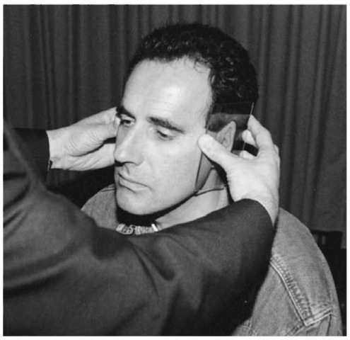
Figure 8 Taking standards (controls) from a subject using a synthetic plate.
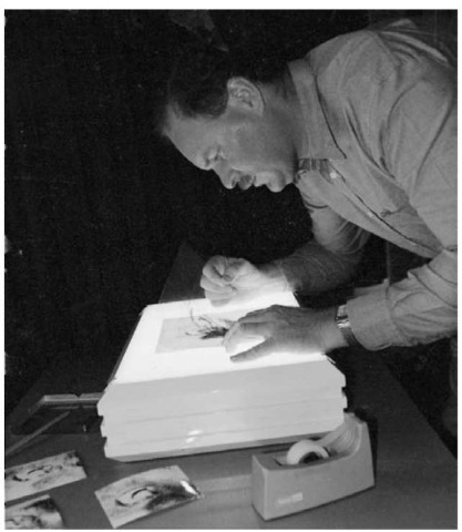
Figure 9 Using the overlay technique for comparison.
(c) Look for the antitragus point and try to cover this point on the unknown with the known.
(d) If these three points more or less fit, look for the antihelix and see whether or not the shape and dimensions are alike.
(e) Look at the lower and upper crura of the antihelix (if present) and see whether or not they fit.
(f) Do the same for the helix rim.
(g) Look at any peculiarities of the helix rim (for instance a tubercle of Darwin) and see whether or not they fit.
If all details more or less coincide you may conclude that the prints are from the same source. If so, continue by:
(h) Attaching the transparency overlay to the photocopy (with a piece of tape).
(i) Cutting both copies into a square, or in case of a resemblance in the skin texture of the cheek, a rectangle.
(j) Attaching both to a piece of paper for presentation purposes.
Step 6 (comparison)
If step 5 was successful, repeat (a) to (h), using the two photocopies of the same trace. The next steps in this procedure are as follows:
(a) Take the ear template, using the stainless steel cross, and put it on top of the prints.
(b) Position the template so that as much as possible of the features of the prints of the ear are cut into two parts.
(c) Remove the stainless steel cross and mark off the inside of the template with a pencil.
(d) Cut the copies along the pencil line, using a roller cutter. A square (10 x 10 cm) will be produced.
(e) Divide the two copies (together) into four parts, according to the indication marks on the top copy (Fig. 10).
(f) Mark the different parts of the copies (known and unknown) carefully with, for instance, a letter and the numbers 1 through 4.
The different parts can be paired and attached to a piece of prepared 10 x 10 cm paper marked with two squares. The new combined images will consist of two different ear prints, but all of the features and dimensions will fit (Fig. 11).
All that remains is the evaluation, deciding in what terms you will express your opinion. It is difficult to give a standard type of report for the comparison of ears and ear prints. Local legal procedures and practices are usually the factors that influence its form and contents.
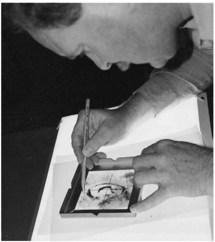
Figure 10 Using the template to divide the photocopy into four parts.
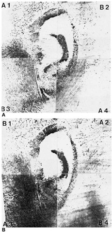
Figure 11 Combining the prints of the known and unknown ears to give (A) A1, B2, B3 and A4; and (B) B1, A2, A3 and B4.
