Introduction
The scientific study of bone and tooth modifications produced by habitual activity patterns, when observable in living and skeletal human subjects, holds an accepted place in the protocol of present-day forensic anthropological investigation. Commonly referred to as markers of occupational stress (MOS), these indicators of one or more persistent and long-term activity-induced changes include musculoskeletal stress markers (MSM) and robusticity markers (RM) in circumstances where ligaments or tendons insert onto the cortical tissue of bone via the perosteum, or where there is a hypertrophy of muscular attachments and thickening of the shafts (diaphyses) of bones of the upper and lower extremities. Some indicators of habitual stress do not involve muscular activity, e.g. the formation of grooves along the enamel occlusal margins of incisor teeth among tailors who hold metal pins in the mouth when sewing. Hair and nails may also reflect an individual’s life history of habitual activities by development of structural irregularities. Thus the term MOS applies to a variety of anatomical modifications and agents. Physiological and cytological processes pertaining to bone remodeling and the biomechanical affects of stress on bone are well documented, particularly in the medical and anatomical literature of sports medicine, industrial medicine, military medicine and orthopedics.
The naming of specific anatomical modifications of skeletal, dental and soft tissue structures as ‘markers of occupational stress’ is attributed to Francesco Ronchese of the Boston University School of Medicine whose text, published in 1948, is entitled Occupational Marks and Other Physical Signs: A Guide to Personal Identification. However, the diagnoses of activity-induced stress began in the Middle Ages with Agricola’s writings on mining hazards and were developed with the advent of the industrial revolution with medical treatises about relief of symptoms suffered by the working classes in farms, factories and trade centers. Anthropologists became interested in MOS from the examination of fossil hominid skeletal remains whereby identifications of squatting facets in Neanderthals and severe occlusal wear patterns of the dentitions of most prehistoric hunting-foraging populations shed light on the behavioral features of extinct populations. The anthropologist who contributed significantly to the study of MOS was the late J. Lawrence Angel of the National Museum of Natural History, Smithsonian Institution, whose investigations were often associated with cases of personal identification brought to him by the Federal Bureau of Investigation (FBI) and other medicolegal agencies. Angel also applied his MOS studies to skeletal series from ancient Aegean and native American populations.
Bone Responses to Stress
In 1892 Julius Wolff, a German anatomist, published his observation that bone tissue has the capacity to increase or decrease its mass in response to mechanical forces. According to Wolff’s Law of Transformation, bone remodeling occurs in well-vasculated subchondrial loci when stress is exerted. The ‘functional pressure’ or load is dissipated by expansion of the bony framework whereby lipping, spurring, exo-stoses and thickening of osseous tissue take place. Severe and prolonged stress is observable macrosco-pically and may be attributed to patterns of habitual and prolonged activity during the life of the individual. Bone resorption occurs in response to persistent compression whereby elevated tubercles and crests are formed by the pull of muscles at the loci of their attachments. However, pull may be related to cortical recession (endosteal growth) when tension results in resorption rather than deposition of bone tissue. Bone remodeling involves both resorptive and depositional functions on periosteal and endosteal surfaces.
Analyses of MOS involve determination of the roles of stress (resistance of bone tissue to the biomechanical action of an external compressional force applied to it), strain (bone deformation or distortion due to tensile or compressive force), shear (a consequence of applied force causing two contiguous portions of a bone to slide relatively to each other in a direction parallel to their plane of contact), and torsion (twisting). These are among the responses of bone tissue to the push and pull of forces exceeding a bone’s elastic limits so that loci of stress do not return to their original form. Excessive stress, strain, shear and torsion may result in destruction (necrosis) of osseous tissue.
Examples of pathological conditions of osteogenesis and physiology of bone tissue, mechanical adaptations of bones and joints, muscular development and function in relation to skeletal morphology and skeletal plasticity are widely documented in the literature. However, there are fewer references to MOS and their significance in reconstructing the lifestyles of extinct populations (when a skeletal record is preserved) or contributing to a positive identification of an individual skeleton. Interest in these latter issues emerged within the last decades of the twentieth century among forensic anthropologists engaged in studies of ancient populations from archeological sites (paleodemography, paleopathology) and as consultants to medicolegal agencies.
Diversity of Markers of Occupational Stress
Recent studies of the causes of bone remodeling at the cellular and molecular levels have led to revision of Wolff’s Law and a more cautious approach to the establishment of direct causality for any specific marker. Factors other than direct response to mechanical loading may be operative, as indicated in the following classification of MOS.
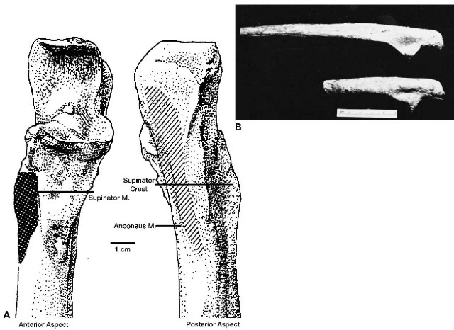
1. The kinds of stressors capable of producing hypertrophy of tendinous or ligamentous periosteal attachments on skeletal organs (enthesopathies and syndesmoses respectively) form irregularities and osteophytes which may be induced by mechanical strain from forces external to the body, as with fractures of the spinous processes of certical vertebrae when heavy loads are carried on the head. Other enthesopathic lesions are induced by internal forces, such as hypertrophy of the supinator crest of the ulna as a consequence of habitual supination and hyperextension of the arm in spear-throwing, ball pitching and use of a slingshot (see Fig. 2).
2. Abrasian occurs when bones are in direct contact at joint surfaces, as is the case in severe osteoar-thritis when intervening structures deteriorate. The joint surfaces exhibit a polished appearance at areas of contact (eburnation).
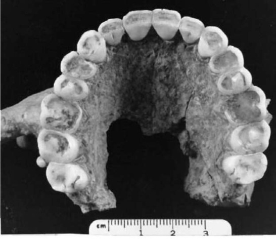
3. Attrition is a condition of wear on enamel, dentine and other dental structures resulting from a variety of habitual behaviors, e.g. tooth grinding, inges-tion of abrasive particles from ambient dust or incorporation of gritty substances in food preparation processes, as well as use of the mouth as an accessory hand when objects are held between the teeth and moved about in the mouth (see Fig. 1).
4. Trauma inflicted on bone and dental tissues may be due to sudden or gradually imposed stress. In cases of bone fracture, osteogenesis originates at some distance from the line of the break in the periosteum and endosteum, then the hiatus is enveloped with replacement of a fibrocartilagen-ous callus. As new bone advances into the callus, irregularity of the area marks it as the original site of trauma. Dental trauma occurs with evulsion, chipping of enamel and displacement of teeth as a consequence of abrupt or gradual stress.

Figure 1 Human maxilla with complete permanent dentition showing severe wear of the anterior teeth. This is attributed to the habitual practice of using the mouth to hold oblects. Specimen is from a prehistoric archaeological site at Mahadaha, India.

Figure 2 (A) Diagrammatic representation of the proximal end of a human right ulna showing hypertrophy of the supinator and anconeous muscle attachments. This may be a consequence of habitual supination and hyperexten-sion of the fore-arm in spear throwing, ball pitching and use of a slingshot.
(B) Hypertrophy of the supinator crest in two human left ulnae from a prehistoric archaeological site at Sarai Nahar Rai, India.
5. Bone degeneration is evident at joints and atrophic patches on adjacent bone surfaces due to loss of volume from osteoporosis, conditions associated with the aging process which are accelerated by obesity, heavy physical labor and a broad spectrum of stressors related to lifestyle. Osteoarthritis is a form of chronic arthropathy characterized by eburnation, cysts, formation of osteophytes, pitting and sclerosis. There is a thin line distinguishing arthritic diseases with an inflammatory condition of degenerative joint disease, traumatic arthritis and osteoarthritis deformans in which little or no soft tissue is involved.
6. Nutritional deficiency not only compromises an individual’s attainment of full ontogenetic development, as reflected in skeletal maturity, growth, body size and stature, but may be attributed to specific irregularities or bone shape (platymeria, platycnemia, platybrachia) and degree of curvature (bowing). The latter condition may not be related to habitual activity patterns in every case since it is related to pathological conditions of treponemal diseases and rickets. Defects of dental enamel, including hypoplasial lines and pits, preserve a record of nutritional stress during the early years of an individual’s life. Analogous markers of episodes of interrupted development are observable radiographically on long bones (Harris lines).
7. In a majority of skeletal samples from different prehistoric and modern cultures, greater muscular-skeletal robusticity is encountered in males than in females, a feature attributed to the harder physical labor in which men engage. Apart from size differences of bones, which may be under the control of genetic and hormonal factors, males, in general, exhibit more robust loci of muscular insertions. Another indicator of sexual difference is the higher frequency of spondylosis and spon-dylolithesis in the vertebrae of male subjects.
8. Racial differences in the incidence of osteoarthritic modifications of knees and pelves between Asian and European populations have been reported, but studies of MOS in ancient and modern populations suggest that cultural differences are more significant than features attributed to ancestry and inheritance. Thus the high incidence of squatting facets on the tibiae of South Asian laborers, when compared to their infrequency in working class people of European descent, is more directly related to differences in resting postures than to genetics.
9. Age differences in expressions of MOS are evident in cases of advanced osteoarthritis in older individuals who had engaged in hard physical labor over long periods of time, but it is not unexpected that any particular MOS could be modified over the course of an individual’s lifetime. There is the potential for early established markers to become less evident with the passage of time under circumstances of discontinuation of the behavior which induces the feature and the action of bone tissue resorption, but this is a subject for future research (Table 1).
Interpretation
As expressions of bone plasticity under pressure from internal and extracorporeal forces, MOS have not been assigned to disorders of metabolism, biochemistry, hormonal and enzymatic imbalances, pathology or neural and vascular disorders. However, certain stress responses, such as those resulting in osteoar-thritis and other threats to the integrity of the body, overlap with classifications of disease. And biochemical and hormonal functions have a role to play in the form and function of bone tissue.
Because experimentation is limited to field and laboratory studies of nonhuman mammals, many MOS have been inferred from historical and archaeological sources, ethnographic accounts and clinical records. Hence interpretations vary in determining ’causes’ of bone and dental irregularities assumed to be responses to habitual activity patterns. Many modern forensic anthropologists support the view that a marker may be attributed to a wide range of cultural practices (not necessarily to a single activity), and that the skeleton registers a mosaic of activities over the course of an individual’s lifetime.
As MOS are interpreted in relationship to the entire skeleton and overall patterns of stress, differences of sex and age become incorporated in the analysis of skeletal remains. Today there is a shift away from the traditional practice of asserting that an irregular morphological feature is the sign of a specific habitual activity towards the keener perception of how bone remodeling takes place, i.e. determining the kinds of modification bone tissue may assume within a broad configuration of behaviors. Accuracy in identification of MOS depends on established standards of observation, availability of well-documented skeletal samples for comparative purposes, classifications of degrees of expression of a real or presumed marker, and other currently accepted approaches to replace the more anecdotal and untestable methods pioneered by earlier investigators. Closer collaboration of forensic anthropologists with biomechanicians, cell and molecular biologists, geneticists, biochemists and physiologists have enhanced recent research efforts in personal identification of human remains and paleo-demographic reconstructions of the lifestyles of ancient populations.
Table 1 Examples of markers of occupational stress reported by forensic anthropologists and anatomists
| Skeletal component | Anatomical structure | Stress factor | Activity |
| Acoustic meatus (ear canal) | Auditory exotosis | Exposure of the ear | Diving to harvest marine foods |
| of the temporal bone | canal to cold water | ||
| Vertebrae | Compression of vertebrae | Vertical compressive | Sledding and tobogganing over rough |
| (‘Snowmobiler’s back’) | force | terrain; snowmobile riding | |
| Fracture of cervical | Hyperflexion of the | Milking a cow when the milker’s head | |
| vertebrae C-6 and C-7 | cervical bodies | is pushed against the animal’s flank | |
| (‘Milker’s neck’) | and the animal shifts position, | ||
| thereby moving the milker’s neck | |||
| Ribs/sternum | Flattening of ribs 6-8 with | Immobility of the | Wearing corsets with rigid stays which |
| curvature of the lower | thoracic cage | are straight and do not permit | |
| sternum | movement of the ribs, as worn by | ||
| women in the eighteenth century | |||
| Ulna | Hypertrophy of the | Supination of the arm | Spear-throwing, pitching a ball, use of |
| supinator crest | sling and atlatl | ||
| Elbow | Lateral epicondylitis of the | Sudden tugging and | Walking a dog on a short leash when |
| joint (‘dog-walker’s | traction on the | the animal is not trained to heel | |
| elbow’) | extended and | ||
| pronated arm | |||
| Thumb | Fracture along transverse or | Fracture | Gripping the saddle horn while flying |
| longitudinal plane of the | off the saddle, as in a rodeo, or while | ||
| diaphysis (‘cowboy’s | riding mechanical bar room bulls | ||
| thumb’) | |||
| Sacrum | Accessory sacroiliac facets | Weight bearing, | Carrying infants or other loads on the |
| at the level of the second | vertical loading in | back over the lumbar-sacral region | |
| posterior sacral foramina | flexion, axial | ||
| and adjacent to the | compression of the | ||
| posterior superior iliac | vertebral column | ||
| spines | |||
| Tibia | Flexion facets at the anterior | Flexion of the knee | Squatting as a resting posture |
| surface of the distal end | |||
| of the tibia at the ankle | |||
| (‘squatting facets’) | |||
| Knee | Iliotibial band of irritation, | Rapid hyperextension | Sharp backward kicking of the leg |
| usually of one leg, leading | of the leg | executed by a team driver at the rear | |
| to osteoarthritic | of a dog sled to spur team to greater | ||
| modifications of the joint | speed over icy and snowy terrain | ||
| (‘Musher’s knee’) | |||
| Teeth | Serrated occlusal surfaces | Wear | Holding and cutting thread by tailors |
| of incisors | and seamstresses | ||
| Anterior tooth loss | Trauma, wear | Using the teeth for power grasping in | |
| holding sled reins or fish lines, or | |||
| results of wrestling or fighting |
