Introduction
A triple murder was committed by two men. The criminals entered through the back door of an appa-rtment and surprised the owner and his friends. After a protracted search of the apartment, they beat, interrogated and eventually killed the victims in succession with large guns. Police arrived at the crime scene and noticed that there was a video surveillance camera similar to those used in a bank. To their surprise the entire event, lasting nearly half an hour from the time the criminals entered the house, was recorded on videotape. Using photo images that were isolated from the tape, the police arrested two suspects. Both suspects challenged the arrest and one of their defense attorneys called in an expert to analyze the tape.
In another incident, a man entered a bank and demanded money. The teller was advised to cooperate and she handed over all the money in her drawer. This event, too, was recorded by a video camera. Police apprehended and arrested a suspect. The terrified teller was shown the suspect and identified him as the man who robbed the bank. All of the other witnesses on the scene disagreed and positively declared that the suspect was not the robber. The suspect, a homeless man, denied committing the crime and insisted he was with a dying friend at the time of the robbery, but the friend, also homeless, could not be located. In this case, too, the defense attorney called in experts to analyze the videotape. In both cases, the prosecution also brought in their forensic experts to ‘prove’ that the suspects matched the criminals in the surveillance tapes.
In the first case, discrepancies demonstrated by video superimposition contributed to a hung jury; in other words, the jury could not agree on a verdict. In the second, a pretrial meeting during which the forensic anthropologists for the defense discussed their findings with the prosecutor led to charges being dropped against the suspect 1 day before the trial was scheduled to begin.
Events such as these are becoming frighteningly common. It is also becoming more common that these crimes are caught on videotape in a commercial establishment or by an ordinary citizen. As more and more surveillance cameras are installed in both public and private venues, the need to develop reliable methods of photographic comparison becomes acute. Methods must not only contain standards of assessment but also be adaptable, because each case presents a unique set of challenges. For one thing, criminals often attempt to disguise themselves with hats, sunglasses and the ever-popular ski mask. Yet the biggest problem is the highly variable nature and quality of surveillance photographs or tapes. In general the quality is poor for many reasons, including film speed, camera distance and angle, lens length and type, and lighting. Another detrimental factor is the reuse of tapes. While this common practice makes economic sense under normal circumstances, the cost can be enormous when it interferes with law enforcement’s ability to identify and convict a criminal. Dark, shadowy, grainy tapes can make identification extremely difficult and can lead to criminals being freed on the basis of ‘reasonable doubt’ if no other evidence exists. A frequent outcome is that the photographic evidence is too poor to be a definitive ‘witness’, and thus this potentially invaluable direct evidence cannot meet the necessary burden of proof. The case must rest on other, often less powerful, circumstantial evidence.
Ironically, it is often the poor quality of the video output that leads prosecutors and defense attorneys to consult a forensic anthropologist. This must be a scientist who is qualified to express expert opinions on many aspects of human biology and can quantitatively and qualitatively analyze and compare images. These can include both skulls and faces in photographs and video footage. Although it seems deceptively simple, this kind of comparison is one of the most difficult to make.
Until fairly recently, cases like this were relatively rare and no standard methodological procedures were in place: investigators simply developed or ‘reinvented’ techniques of their own to fit the situation. The purpose of this article is therefore to discuss technical and methodological problems, new research and development, and current methodology that has been used with repeatable success in comparing facial images. These approaches include facial morphology, photoanthropometry and video superimposition. Research, experience, cases studies and trial work have demonstrated that these approaches can be applied to reach a conclusion, be it positive, negative or indeterminate. It has also brought to light the weaknesses inherent in this kind of analysis and sparked further research in the field.
Technical Considerations
When consulting on any case of this nature, it is vital to ascertain the source of the photographic evidence. It is always preferable to use the originals because even a good copy will lose some detail. Law enforcement agencies often make enlargements or enhance the original to bring out detail and facilitate finding a suspect at the beginning of the investigation. However, during the process of image analysis, an enhanced photograph should not be used for comparison or superimposition because the processing can result in distortion. This was evident in a case where the spotlights in the original were round, while the same fixtures appeared oval in the enhanced still. Obviously, the facial contours and features would be similarly affected.
Many aspects of the equipment itself should be considered, such as the distance and angle of the surveillance camera with that of the subject being photographed. The type of lens in the camera can also affect perspective, as with a very wide-angle or fish-eye lens that is sometimes used to cover a large area. In general, an image may be elongated if the subject-camera distance is short. Greater distance might make a face look rounder. Another aspect one has to consider is the angle of the images. In a full frontal view, angulation would affect the height of the face and features like the nose. A face positioned near the standard Frankfort horizontal plane would be longer than one tilted upward or downward. In profile, the length of the nose or face itself is not affected by head tilt. While facial width dimensions would generally not be altered by moderate upor down tilt, they are sensitive to even slight lateral deviations.
Care must also be taken in the choice of photographs used for comparisons that have been provided by the family of the individual in question. If they were done professionally, there is always the possibility that the prints were retouched. This, of course, would make them inappropriate for a comparison because the purpose of retouching is to improve appearance by eliminating defects or softening imperfect features. In addition, even makeupcan interfere with the process by obscuring scars or creating a younger look. Ironically, it is just these characteristics that can be the all important factors of individualization which are necessary for positive identification.
Morphologic Approach
The human face is a reflection of the individual uniqueness of a person. There can be hints to many aspects of the persona – everything from personality and temperament to overall health and levels of stress. Biologically, the facial phenotype is a product of genetics and environment that reflects features of populations in specific regions. Local populations are products of long-term inbreeding within the community as well as with nearby neighbors. To exemplify this, the so-called central European Alpines are described as basic round-headed Caucasoid brunettes with medium width noses. Their primary sorting criteria include dark to medium brown hair and eye color, globular heads with high foreheads, a mesor-rhine nose with a slightly concave or straight profile and fleshy, ‘blobby’ and often elevated tip. They also have olive (brunette) white skin color, abundant hair and beard, round or square face with prominent gonial angles. Although such persons may now be seen on every continent, they will be much more likely to be indigenous to Europe and their frequency is expected to be higher in that region.
During the nineteenth and twentieth centuries, scientists developed remarkable research techniques for analyzing human facial variation. Practically every detail of the face has been systematically divided into gradations and categories based on relative size, shape, presence or absence. In addition, some of the more complex facial features were simplified to allow more objective evaluations. For example, the facial profile can be assorted by projection, e.g. mid-face jutting with nose protruding, as opposed to chin or forehead jutting, and midface concave. Full face outlines can also have many variations, such as elliptical, round, oval, square or double concave around the zygomatic region, among others. Table 1 shows some of the commonly observed facial features and their respective scales of observation. While the table was originally developed to observe a living person, some of the same observations can be made on prints captured from video or movie footage, and photographs. The evaluation of the face and its classification is not a simple matter and requires considerable experience of anatomy and understanding of human variation. The facial features listed in the table also provide an opportunity for the investigator to systematically examine the entire face in detail.
The human face is a dynamic structure and can transmit a wide range of expressions, from very minute to greatly exaggerated. Even the most subtle changes in expression may create a different perception in others. Emotions such as happiness, curiosity, concern, anger, fear, worry or surprise are instantly recorded and can just as quickly disappear. Physical and more permanent alterations result from aging, disease, weight gain/loss, graying and loss of hair or beard, and exposure to the sun. Studies have reported that smoking can accelerate wrinkling. Over a lifetime, all these external and internal factors leave their impressions and create a nearly infinite range of individual variation, both within and between people. These become important factors, especially when comparing images taken over time.
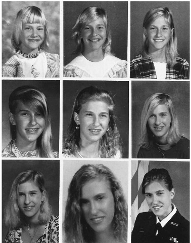
During growth, facial features change noticeably. In the face they include changes that result from the enlargement of the head, nose, jaws and eruption of teeth. Figure 1 shows physiognomic changes from midchildhood (6.5 years) through late adolescence (18 years). Most obvious is the elongation of the lower half of the face and nose. Yet the pointed chin shape remains consistent. This stems from the fact that the bony foundation of this region must be established early in life to support the development and eruption of the deciduous and permanent dentition. The incisors (and permanent first molars) are the first adult skeletal components to form in the body, and most of the permanent anterior teeth are either well in progress or completely erupted by about the age of 7 years. Not only does this affect the overall height of the face, but research has demonstrated that the mandibular symphysis takes adult shape at about that time, and from then on merely increases in size. Eyebrow form and density shows little variation throughout this 12 year period. It is to be expected that males would developbushier brows during adolescence, as well as a beard. There were no obvious new folds and creases in the face that can be associated with growth. The forehead height and the location and shape of the hairline also appeared unchanged. Ear forms are clearly visible in several pictures and the ear lobe is attached. The picture at age 17, in the middle of the bottom row, differs from others as it shows some retouching and smoothing of creases. Hair form also remained straight during this time, even though the retouched image shows a curly hair style. In this case the change resulted from a perm, but it is not unusual for hair form to change during adolescence (e.g. a straight-haired child can become curly, or vice versa). In any event, hair style is too easy to alter to be used as a primary criterion for photographic comparison. Actual form, texture and color can only be determined by microscopic analysis of the hair itself.
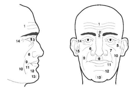
It is an indisputable fact that skin develops wrinkles with age. However, extreme variability in onset and progression makes it impossible to quantify the rela-tionshipof wrinkling patterns to age. It has always been a popular assumption that the chin and the eyes are the first places to look for the presence of wrinkling. Figure 2 shows wrinkle and fold patterns in a face. As noted above, causative factors are numerous. The most obvious changes are the formation of new lines and the deepening of furrows. Subtle changes include asymmetry of the eyelids, which occurs when one loses its elasticity earlier than the other. This loss of tone often gives the impression of an epicanthic fold in a Caucasoid. Another factor is that cartilage can grow throughout life, leading to lengthening of the nose and enlargement of the ears. This can be particularly troublesome when attempting to compare a photograph of a man in his twenties with his own picture some 40 years later.
A primary focus of facial identification research is to isolate features that can be considered as factors of individualization. Such features may not be very easy to detect, especially in a relatively closed population where interbreeding is the norm. In that situation, many individuals share similar features. It is also true that, while populations share some obvious attributes, everyone has features that make him or her distinctive. It is these features that we rely upon in our everyday lives to recognize each other, even though we may not be consciously aware of the process. Even children will embarrassingly blurt out that someone has a long nose, big mouth or ears that ‘stick out’. It is the scientist’s job to isolate and identify even the most subtle differences in size, shape, asymmetry and proportion.
Table 1 Morphological characteristics of the head and face observable on photographs and living persons
| Facial forms | Baldness | Eyebrow thickness |
| Elliptical | Absent | Slight |
| Round | Slight | Small |
| Oval | Advanced | Average |
| Pentagonal | Complete | Large |
| Rhomboid | Beard quantity | Concurrency |
| Square | Very little | Absent |
| Trapezoid | Small | Slight |
| Wedge-shaped | Average | Average |
| Double concave | Hairy | Continuous |
| Asymmetrical | Hair color: Head and beard | Eyebrow shape |
| Facial profiles | Black | Straight |
| Jutting | Brown | Wavy |
| Forward curving | Red bright | Arched |
| Vertical | Golden | Eyebrow density |
| Concave | Red | Sparse |
| Lower jutting | Grey | Thick |
| Upper jutting | White | Bushy |
| Forehead height | Red pigment | Nasion depression |
| Low | Absent | Trace |
| MediumPresent | Slight | |
| High | Iris color | Average |
| Forehead width | Black | Deep |
| Small | Brown | Very Deep |
| MediumGreen-brown | Bony profile | |
| Broad | Blue-brown | Straight |
| Skin color | Green | Concave |
| Pale | Grey | Wavy |
| Brunette | Blue | Convex |
| Brown | Other | Bridge height |
| Chocolate | Eyefolds | Small |
| Black | Absent | Medium |
| Vascularity | Internal | High |
| Slight | Slight | Bridge breadth |
| Average | Average | Very small |
| Extreme | Developed | Small |
| Freckles | Median | Medium |
| None | Slight | Large |
| Few | Average | Tip thickness |
| Moderate | Developed | Very small |
| Extreme | External | Small |
| Moles | Slight | Average |
| None | Average | Thick |
| Few | Developed | Tip shape |
| Moderate | Palpebral slit | Pointed |
| Extreme | Down | Bilobed |
| Hair formHorizontal | Angular | |
| Straight | Up slight | Rounded |
| Low waves | Up moderate | Blobby |
| Deep waves | Up extreme | Snub |
| Curly | Opening height | Septumtilt |
| Frizzy | Small | Upward |
| Woolly | MediumUp slightly | |
| Texture | Large | Horizontal |
| Fine | Upper lid | Down slightly |
| Moderate | Low | Downward |
| Coarse | Medium | |
| Wiry | High | |
| Nostril formUpper lip notch | Ear size | |
| Slit | Absent | Small |
| Ellipse | Wavy | Medium |
| Intermediate | V-Shape | Large |
| Round | Mouth Corner | Ear projection |
| Nostril visibility | Straight | Slight |
| Lateral | Upturn | Medium |
| None | Downturn | Large |
| Slight | Alveolar prognathismHelix | |
| MediumAbsent | Flat | |
| Visible | Slight | Slight roll |
| Frontal | MediumAverage | |
| None | Pronounced | Very rolled |
| Slight | Malars | Anti-helix |
| MediumAnterior projection | Slight | |
| Visible | Absent | Medium |
| Nasal alae | Slight | Developed |
| Compressed | Medium | Darwin’s point |
| Slight | Pronounced | Absent |
| Flaring | Lateral projection | Present |
| Extended | Compressed | Lobe |
| Lip thickness | Slight | None |
| Very thin | MediumSoldered | |
| Thin | Pronounced | Attached |
| Average | Chin projection | Free |
| Thick | Negative | Long and free |
| Eversion | Neutral | Other features |
| Slight | Slight | Birth marks: |
| Small | Average | |
| Average | Pronounced | Moles: |
| Everted | Chin type | |
| Lip seamMedian | Wrinkles: | |
| Absent | Triangle | |
| Slight | Bilateral | Asymmetry |
| Average | Chin fromfront | |
| Present | Small and round | Fatness: |
| Integument lips | Wide and round | |
| Thin | Pointed | Mustache: |
| Average | Chin shape | |
| Thick | Dimple | Beard |
| Philtrumsize | Cleft | |
| Small | Double chin | Sideburns |
| Wide | Gonial eversion | |
| Philtrum shape | Compressed | Trauma: |
| Flat | Slight | |
| Deep | Moderate | Surgery: |
| Sides parallel | Everted | |
| Sides divergent | Very Everted | Scars: |
| Glasses: | ||

Figure 1 Pictures depicting physiognomic changes from middle childhood through late adolescence. Top row: ages are 6.5, 9.5 and 10.5 years; middle row: 12.5, 13.5 and 14.5 years; and bottom row: 15.5, 17 and 18 years.

Figure 2 Wrinkle patterns: 1, horizontal forehead wrinkles; 2, vertical wrinkles of the glabella; 3, wrinkles of the nasal root; 4, eye fold below the orbit; 5, eye-cheek fold; 6, nose-cheek wrinkle; 7, nose-lip fold; 8, nose-lip fold; 9, cheek-chin wrinkle; 10, cheek-chin fold; 11, mouth corner fold; 12, lip-chin fold; 13, chin cleft; 14, temporal wrinkles; 15, ear wrinkles.
Every feature must be analyzed, for each gives clues at both the population and individual level. Hair color or form is important in race assessment. While Cau-casoids can have nearly every color, texture and form, Mongoloids are characterized by straight, coarse dark hair and Negroids by dark, frizzy hair. Although hair can be easily colored, curled or straightened, the genetically programmed characteristics can be determined by microscopic analysis. Certain features point to populations within a given racial phenotype. Asian Indians, for example, are Caucasoid, but form a distinctly recognizable group. After sex has been determined, categorization of racial affinity is of primary importance. There must be sound scientific reasoning underlying the selection of features that are unique and unvarying enough to eliminate certain groups, and eventually individuals, as candidates. Attributes such as nose form, lipthickness and alveolar prog-nathism are among those structures known to show significant variation between races and are thus good structures with which to begin an analysis.
Admixture is increasingly common and can be both problematic and individualizing at the same time. Since the general public assigns racial affiliation based on the most dominant visible (phenotypic) characteristics, admixture may not always be apparent to casual observers and care must be taken not to eliminate candidates for identification. On the other hand, admixed features can be factors of individuali-zation, such as an ostensibly black man with an unusually long narrow nose and little prognathism.
There are a number of considerations that should govern the choice of comparative sites. Often the selection is dictated by what is visible or clearest in the images. Examples of recommended sites include the eye, especially details like interpupillary distance,nasion or glabella, the tipof the nose, the base of the chin, ear shape and type of protrusion (upper, lower or both). It is advisable to choose structures that are most resistant to the ravages of time, cosmetic modification and weight changes. Easily modified features, especially those relating to hair (length and color, sideburns, beards and other facial hair), should be avoided. Even the hairline often recedes and changes shape during the adult lifespan.
In a recent attempt to classify human faces, 50 sets of photographs (full, profile and three-quarter views) of white male adults were analyzed. Each set was examined using 39 facial features selected and modified from Table 1. The researchers found that height and width dimensions, not defined by fixed points and requiring judgment by the observer, to be the most unreliable and unpredictable. Examples include forehead height and hair length. Features like face shape (e.g. oval or triangular) had higher rates of interobserver agreement, and pronounced ear projection was found to be one of the best discriminators.
Photoanthropometry
A second approach is based on the size and proportionality of various facial features. The technique traces its roots to traditional anthropometric methods. For the sake of clarity, it is referred as photo-anthropometry. These quantitative approaches diverge when it comes to what is being assessed. Anthropometry is based on measurement of the living and deals with a three-dimensional structure. In the living, landmarks delineating measurements can be palpated and located precisely and repeatably with experience. When photographs are involved, the process becomes essentially two-dimensional. Photographic quality affecting the distinctness of lines and borders as well as the problems of perspective and depth of field are major factors that increase the difficulty of this analysis and lower the level of precision that can be expected. In addition, the forensic expert can only control the photographs he or she takes for comparison and, no matter how good those are, the limiting factor is the quality of the evidentiary photograph. Although the developing process can yield a brighter print or increase contrast, as noted above, care must be taken to avoid enhancements that cause distortions in size or shape.
A key element in the use of photoanthropometry is the formulation of indices based on proportions rather than absolute size. They assess the relationship of one structure (e.g. nose length) to another (e.g. total facial height). As in all analyses, the landmarks used for indices must be clearly visible and defined if they are not standard sites. An important consideration for this approach is not to limit an analysis to preset or traditional landmarks. The quality and angulation of the image may dictate the use of unusual points that can be clearly defined and repeatably located on both images. Consistent replicability is essential, especially for courtroom presentations. This can be achieved by using a clear overlay to mark the reference points on each photograph without altering the evidence, obscuring features or interfering with other kinds of analyses.
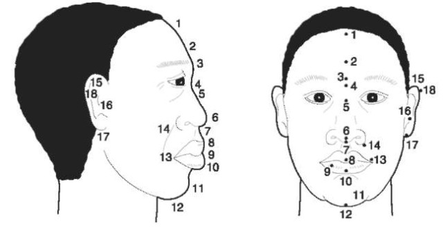
To begin a metric photographic analysis the pictures should be copied and enlarged to approximate the actual size of the faces if possible. However, enlargements should only be made if they do not compromise the quality of the image. Once the landmarks have been chosen, they must be marked with a pen with a very fine tip on both photographs (but never on originals) to be compared. It is absolutely essential not to alter the originals. Suggested sites visible (in full face, profile or both) when the picture is oriented in the Frankfort horizontal plane are illustrated in Figure 3 and are described as follows:
1. Trichion: midpoint of the hairline (full face and profile).
2. Metopion: most anterior point of the forehead (profile only).
3. Glabella: midpoint between the eyebrows on the median plane (profile only).
4. Nasion: deepest point of the nasal root (profile only).
5. Midnasal point: midpoint between the endo-canthions (full face only).
6. Pronasale: most anterior point of the nose tip (profile only).
7. Subnasale: point where the nasal septum meets the philtrum (can be located in both full and profile if the nose tipis horizontal or elevated).
8. Superior labiale: midpoint of the vermilion seam of the upper lip (full face and profile).
9. Stomion: midpoint of the occlusal line between the lips (full face and profile).
10. Inferior labiale: midpoint of the vermilion seam of the lower lip(full face and profile).
11. Pogonion: most anterior point of the chin (profile only).
12. Gnathion: Most inferior point of the chin (full face and profile).
13. Cheilion: corner of the mouth (full face and profile).
14. Alare: most lateral point of the nasal wings (full face and profile).
15. Superaurale: most superior point of the ear (full face and profile).
16. Tragion: most anterior point of the tragus (profile only).
17. Subaurale: most inferior point of the ear (full face and profile).
18. Postaurale: most posterior point of the ear (profile only).

Figure 3 Anatomic landmarks of the face: 1, trichion; 2, metopion; 3, glabella; 4, nasion; 5, midnasal point; 6, pronasale; 7, subnasale; 8, superior labiale; 9, stomion; 10, inferior labiale; 11, pogonion; 12, gnathion; 13, cheilion; 14, alare; 15, superaurale; 16, tragion; 17, subaurale; 18, postaurale.
Many measurements can be taken from these two views. If trichion (1) is clear, it can be the starting point for the majority of vertical measurements, e.g. 1-4 (1-5), 1-7 (or 1-6), 1-8, 1-9, 1-10,1-12, 14-14 and 16-16. If trichion is not available or the hairline is receding, then 4 (or 5) can be the starting point. Facial width measurements can cover 1-16, 2-16, 416, 6-16, 7-16 and 15-17.
As mentioned earlier, the examiner is not limited to preset landmarks and others can be defined and used if they are better adapted to the images in question and are clearly visible in both photographs. Because of the variability in the type and quality of evidence each case presents, a standard set of dimensions cannot be established. All measurements must be taken with a precision caliper with vernier accurate to at least one decimal place.
Finally, since absolute size is not reliable without a scale, indices must be calculated from these measurements to insure that the values are comparable. Although standard-sized objects may be in the photograph and thus can be used as a scale, this rarely happens. In most cases it is impossible to determine the actual dimensions of the face and its features. Approximations are not recommended because the potential for error is too great. By relying on relative proportions, the index functions to eliminate the incomparability resulting from absolute size differences between images. An index is created as follows:

For linear dimensions, it is best to use the maximum dimension as a constant denominator (e.g. 4-7/1-12, 4-8/1-12, 4-8/1-12, etc.). Width or breadth can also be assessed in relation to height or other facial dimensions. Computer spreadsheets are recommended for this purpose because they can quickly and accurately generate the desired values.
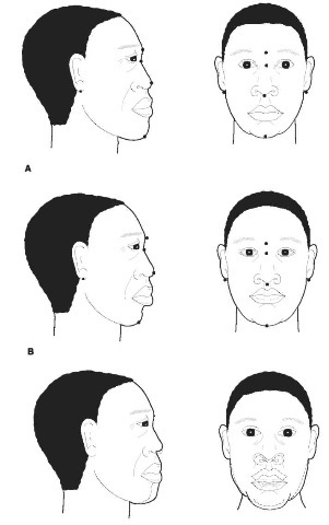
This metric procedure is demonstrated using persons A and B in Figure 4. Table 2 presents the values measured from the original drawings. Three measurements were taken for this exercise from both full and profile views. Since the actual sizes of these (fictitious) people are not known, three indices per facial view were calculated for this demonstration, but additional measurements can always be taken and indices computed to provide support for differences or similarities. The only restriction is that the points be clear on both pictures. It is obvious that person A has definite anthropometric differences from person B. Person B has a longer nose in relation to both facial height and breadth. He also has a wider face in relation to facial height. This was the case in both the frontal and profile images.
The marked photographs must be used for the investigation and submitted to the court as evidence. Copies should be made by reshooting prints after the photographs have been marked. This is the only way to duplicate exactly the positions of the marks. Photocopies should not be used for official purposes.
The aim of photoanthropometry is to compare metrically the proportional relationships of one photograph to another, rather than assess absolute visual differences and similarities as in morphological comparisons. While replicable quantification may reduce the subjectivity of photographic comparisons, one still cannot always reach a definitive conclusion regarding the matching of images. The most obvious problems are:
• The photographs are usually taken under different conditions.
• Proportional aberrations can be caused by differences in the lens angle and the camera-to-subject distance.
• Some photographs may have been retouched so that facial features may not match those of the unaltered photographs, even if it is the same individual.
• Weight and age differences can change the location of landmarks.
• Differences in facial expression may create differences in certain measurements (e.g. mouth size). This is especially true when a smiling visage is compared with a frowning one. The alterations of the facial musculature during laughter, for example, may widen the mouth, shorten nose-to-mouth distance and lengthen the face.

Figure 4 Profile (left) and frontal (right) sketches of fictitious persons A (top row) and B (middle row) for photoanthropometric comparison using marked points. The bottom row shows some of the discrepancies in the nose length, mouth position and lower facial border that would be seen when the faces are superimposed: —person A; — person B.
Table 2 Comparison of indices calculated from the dimensions of frontal and profile views of person A (unknown) with person B (suspect)
| Dimensions | A | B | Indices | A | B |
| Frontal view | |||||
| Glabella-Menton (face h) | 39.4 | 36.1 | Nose h/face h | 34.3 | 38.8 |
| Nasion-Subnasale (nose h) | 13.5 | 14.0 | Nose h/face w | 36.4 | 38.1 |
| Subaurale-Subaurale (face w) | 37.1 | 36.7 | Face w/face h | 94.2 | 101.7 |
| Profile view | |||||
| Glabella-Menton (profile face h) | 40.0 | 37.6 | Nose h/face h | 28.0 | 32.2 |
| Nasion-Pronasale (profile nose h) | 11.2 | 12.1 | Nose h/face w | 34.4 | 38.5 |
| Subaurale-Pronasale (profile face w) | 32.6 | 31.4 | Face w/face h | 81.5 | 83.5 |
Further research in photographic analysis is needed to developmethods that can account for mensural variations, arising from differences in pose and angle, by experimenting with control subjects. Studies should follow exacting scientific protocols, including rigorous tests of empirical data. Experimentation along these lines has already begun.
Photographic Video Superimposition
Photographic superimposition is the method whereby two comparably enlarged photographs are superimposed using video cameras, a mixer and a monitor. Current video technology can eliminate the laborious work and expense of photographic enlargement and the difficulties of object orientation (e.g. a living person or a skull) to duplicate the pose of the picture.
From a technical standpoint, the procedure can be readily accomplished with the right equipment. The challenging part is judging the quality of fit, identifying inconsistencies and knowing the range of acceptable variability.
The aim of this approach is to determine if facial features and dimensions match when two images are superimposed. Following proper placement of the cameras, photographic distances are adjusted to make the images the same size. At least three points must be aligned. These should include fixed points like the eyes, when visible, and one other clear recognizable part, like the nose, jaw line or contour of the head. A mixer then allows several types of visual comparison. In a ‘vertical wipe’ one face passes laterally across the other. The wipe can also be horizontal from topto bottom, and vice versa. A ‘fade’ makes one face disappear into another, with the second image eventually replacing the first. A spot analysis can be used to emphasize a selected region of the face. The spotlight can be focused on a particularly telling feature to call attention to differences or similarities. This should always be used to highlight an area of discrepancy, such as when the chin of one face extends beyond that of the other.
Fig. 4 (bottom row) illustrates the process of super-imposition. The dotted lines (representing person B) highlight some of the discrepancies in the nose, mouth and lower face between person A (solid lines) and person B. It can be clearly seen that person A has a shorter nose, more protruding chin and narrower face.
As can be expected, there are a number of problems associated with superimposition, many of which also plague morphological and anthropometric comparisons. Of all types of superimposition, photo-to-photo is the least accurate. A problem unique to this comparison is the near impossibility of finding photographs that are taken under exactly the same conditions and with identical poses and expressions. The greatest accuracy can be achieved by superimposing a photograph on a living person or a skull, i.e.three-dimensional objects. This allows the best possible alignment because the latter are not in a fixed position and can be easily adjusted to the orientation of the photograph. Another advantage is that the living person can imitate the facial expression in the photograph.
New applications of computer digitization have the potential to offer a more objective mode of superim-position. A computer can be programmed to quantify and evaluate the characteristics that differentiate one face from another. A program has been developed that digitizes projective symmetry in photographs and uses the resultant patterns to assess the comparability of two individuals. A composite image combining sections of each photograph is generated and if the components from both pictures fit perfectly, it is likely that the images are those of the same person. The developers report success even when a disguise is used. The latest concept in this field is experimentation with the application of a neural network system to compare two images. The aim is greater objectivity and the elimination of biases that may arise from individual observations. It also attempts to reduce the heavy reliance on symmetry that is the focus of digitization programs. This study even claims that age-related changes may be detectable with this technique. As with all new technology, rigorous independent testing is necessary before final judgments of effectiveness can be made.
For now, the best that can be said about technological advances concerns the very positive benefits derived from the use of video and computer systems that allow the expert to develop and demonstrate a series of visual effects and focus on morphological details. This in turn enables interested parties, like jurors, attorneys and judges, to see the entire procedure and visualize exactly how the experts reached their conclusions.
Expert Testimony
Being an expert witness is an awesome responsibility fraught with pitfalls that can have serious consequences. It is hard enough to carry out this job in areas with well-established criteria for judging evidence. Thus, the professionalism and experience of the witness becomes paramount in a developing field like this.
When an expert opinion is requested, the qualified scientist must demonstrate in detail how the analysis, superimposition for example, was carried out. The best approach is to allow plenty of time to show and explain how one photograph is superimposed over another. In most cases superimpositions do not match exactly, although a very slow fade or rapid wipe may give the illusion of a perfect match as one image is almost imperceptibly replaced by the other. Anything that can obfuscate inconsistencies must be avoided to maintain the integrity of the procedure. Following a complete initial wipe, the process of dissolving one image into the next should be repeated with pauses to focus on areas (highlighted by spot analysis if called for) where there are marked differences or excellent concordance. The expert should literally point out and trace these areas on the screen during pretrial deposition or testimony in court to insure that there is no misunderstanding.
An important aspect of being an expert witness is that one must pay scrupulous attention to any presentation by opposing experts. An expert must be prepared to advise counsel of errors or attempts to mislead. In some cases opponents focus only on what fits or try to distract by showing a nice overall agreement. For example, some witnesses draw attention to agreements in ear or nose length while ignoring obvious discrepancies in the chin and forehead. There are stringent requirements (addressed below) for declaring a positive match. An expert must not accept identification based solely on agreement in a few general features like facial roundness, projecting ears and receding hairline. One must also not lose sight of differences in expression between images such as an open mouth that is dissolving into a closed one or a smile that ‘disappears’. Camera ‘tricks’ can either enhance or minimize variation between pictures. That is why we emphasize again that everything must be clearly described – what fits and what does not – no matter which side you represent. The same, of course, applies to metric and morphologic analyses. It can only hurt an expert’s case and credibility to have the other side point out something that he or she failed to mention or attempted to obscure.
What conclusions are possible from photographic comparisons? Under optimal conditions an expert can make a positive identification. A definite declaration of positive identification is possible only if the following conditions are met: (1) all points match exactly with no discrepancy; (2) the images are clear and sharp at all points of comparison; and (3) there is a factor of individualization visible on both images. The third and absolutely essential feature can be a scar, gap between incisors, asymmetry, oddly shaped nose or head or other rare or unusual attribute that sets this individual apart. A match can also be ruled out. This conclusion can be reached when there are obvious discrepancies in the size or shape of attributes between the photographs. Similar-looking individuals can also be definitively differentiated by, for example, the presence of a scar in one image, but not the other. In these situations, the expert can confidently state that the suspect is not the person photographed committing the crime. Lastly, most conclusions fall into the inconclusive or indeterminate category. This simply states that the expert cannot determine with 100% certainty that there is or is not a match. This situation can occur for a number of reasons, such as when there is a good match between the images but there is no factor of individualization. Although human variation is almost infinite, many people share certain features that are similar. Reaching definite conclusions is a real problem when someone has so-called ‘average’ features. This designation can also apply when one of the images is not very clear or sharp. In the authors’ experience, the question of ‘how possible?’ often arises. In this situation, experts can use their discretion to indicate a stronger or weaker association, but there are no statistics on this type of probability, and no attempt should be made to guess at a ‘ball park’ figure. Most importantly, and as with any scientific pursuit, restraint must be exercised not to make any pronouncements that cannot be solidly supported by the data.
Discussion and Conclusions
The global rise in crime and the resultant video technology applied to fight it have created a growing need for anthropological expertise in image analysis. The three methods described in this article have been used in legal proceedings to compare one photograph with another (or videotape frame). They are: (1) morphological – detailed comparison of facial features following the form in Table 1; (2) photoanthropometry – quantitative analysis based on measurements of facial dimensions and the generation of indices based on them; and (3) photographic video super-imposition – superimposition of one photograph or videotape frame over another. While it is rare to be able to make a positive pronouncement, two or more of these should be combined whenever possible to build a strongly supportive foundation for the final conclusion.
Of the comparative techniques presented here, the morphologic approach offers the best chance of obtaining a positive identification or ruling out a match, as is the case in skeletal analysis. It is the assessment of visible morphologic features that ferrets out factors of individualization, when present. Unfortunately for the investigator, many people simply do not have noticeable disfigurations or unusual facial elements. Most folks are ‘average looking’ and they inevitably fall into the ‘indeterminate’ category. Although the general public and experts alike clearly notice that people look different, they are often at a loss to describe exactly how two similar individuals differ.
There is a vague sense that maybe one is a bit ‘prettier’ or more ‘masculine’. What makes one individual different from all others is the highly varied complexity of many features and how they come together. It cannot be overstressed that a match must not be made on the basis of a general look, i.e. the common traits that define a racial phenotype, for example. Interpretations and conclusions must always be made with caution.
Photo-to-photo superimposition can provide an easy comparison of images and show discrepancies. It brings together two separate images and illustrates the proportional relationship of facial features in both photographs. Even if differences are distinct morphologically, or if there are clear factors of individualization, a superimposition may still show the location of these individualizing factors in the two images.
The methods discussed here do have the potential to conclude that there is a positive match, or rule one out decisively. However, it cannot be overemphasized that, no matter how good our methodology becomes, the process of comparison and superimposition is seriously compromised by the often extremely poor quality of surveillance photographs. In the absence of a skull or live subject that can be posed, the less than ideal condition of pictures representing missing persons further compounds the difficulties of analysis. This makes it difficult, if not impossible, to precisely place landmarks and borders for comparison. Another obstacle is the difficulty of determining absolute size. The probability of error is too great when one must extrapolate true dimensions in a photo with no scale. An invaluable improvement would be the installation of a measuring scale in the camera that would appear on each frame of the film. With the size factor accounted for, it would be possible to rule out many suspects with a few, simple, raw metric comparisons.
Finally, at the present level of development of this emerging specialty, conclusions in most cases will remain possible or indeterminate, at best. Therefore, photographic evidence often cannot be the decisive factor upon which to convict or exonerate a suspect. Technical and methodological difficulties must both be addressed for significant progress to be made in this type of identification. The expert witness must be thoughtful and responsible in making conclusions. Stretching the data to the point of conjecture must be avoided, and murky or doubtful areas presented as such. Credibility and cases can be lost unless experts fully comprehend and are able to communicate both the potential and limitations inherent in photographic identification.
