The Pelvic Girdle
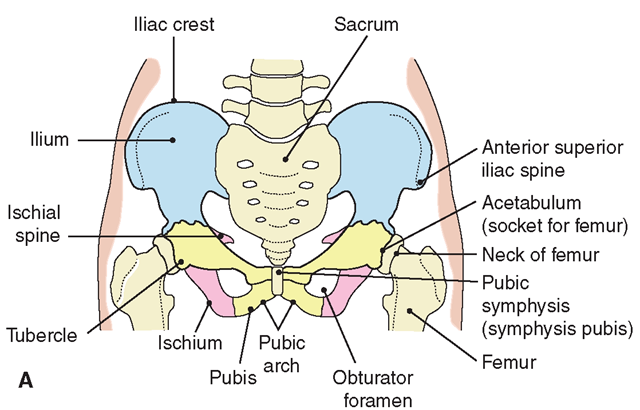
Two large, irregularly shaped innominate hip bones (os coxae) attach posteriorly to the sacrum (Fig. 18-15) to form the pelvic girdle or pelvis.
FIGURE 18-15 · The pelvic bones. (A) Anterior view. (B) Lateral view.
The pelvis supports the spinal column and protects parts of the digestive, urinary, and reproductive systems. The pelvic bones spread outward at the top and become narrow at their front lower edges. The ilium is the upper flaring portion of the pelvis that is usually identified as the hip bone. The ischium is the lower, stronger portion. The pubic bones meet in front and are joined by a pad of cartilage; this juncture is called the symphysis pubis. Connected to the sacrum and coccyx posteriorly, these bones form the pelvic cavity, which houses the urinary bladder, the rectum, and in women, the uterus and ovaries. In men, the prostate and some glands are within the pelvis. A woman’s pelvis is larger and wider than a man’s, which provides room for development and vaginal delivery of the fetus. The angle of the pelvic opening (pubic arch) is less than 90 degrees in men and greater than 90 degrees in women.
Special Considerations : LIFESPAN
In the fetus, pelvic bones develop as three separate bones known as the ilium, ischium, and pubis, which usually fuse by the time growth is completed. The age for normal completion of bone growth varies, but is usually complete by age 25.
NCLEX Alert The NCLEX not only requires knowledge of the structures and functions of muscles and bones, it also requires that you know the differences of nursing care between injuries, illness, or inherited disorders of muscles or bones.
THE MUSCLES
Functions of the skeleton include giving the body its shape and providing mobility, using bones as levers. Without the aid of muscles, the body would be immobile. Although the skeleton determines the size of the body’s framework, muscle and fat determine body shape. Functions of muscles are body movement, blood circulation, and heat production (Box 18-3). All muscle tissues have specific characteristics (contractility, extensibility, elasticity, and irritability), as described later in this topic.
BOX 18-3.
Functions of Muscles
Voluntary Movement
♦ Enables walking, standing, sitting, and other movements
♦ Maintains body in upright position
♦ Participates in body balance
Involuntary Muscle Action
♦ Maintains heartbeat to pump blood
♦ Provides arterial blood flow
♦ Promotes lymphatic and venous blood return to heart
♦ Dilates and contracts blood vessels to control blood flow
♦ Maintains respiration
♦ Performs digestion processes
♦ Performs elimination processes
♦ Participates in reflexes
♦ Enables all other involuntary actions of body
Protection
♦ Protects body in emergency by reflex action
♦ Covers, surrounds, and protects internal organs (viscera)
♦ Supports internal organs
Miscellaneous
♦ Produces heat
♦ Assists in maintaining stable body temperature (in shivering, “goose flesh," muscles give off heat)
♦ Provides shape to body
Muscle Classification
Three types of muscle tissue by function: skeletal, smooth, and cardiac. Each is also identified according to appearance. Skeletal and cardiac muscles are striated;they consist of fibers marked by bands crossing them, giving them a striped appearance. Smooth muscle is nonstriated. Muscles are further determined to be voluntary or involuntary. Muscle tissue is approximately 15% denser than adipose (fat) tissue. Table 18-4 presents the different muscle types.
Skeletal Muscles
Skeletal muscles, which control movements of the skeleton, are under voluntary (conscious) control. There are more than 630 skeletal muscles in the human body, constituting approximately 40% of body weight (Fig. 18-16). Their functions include locomotion, facial expression, and posture. The two types of voluntary muscles are fast-twitch—those which contract quickly and powerfully, but encounter rapid fatigue, and slow-twitch—those which can sustain a contraction, but do not exert great force. It is believed that people are born with a predisposition toward more fast-twitch or more slow-twitch muscles.
Key Concept The masseter (jaw) muscle is considered to be the strongest muscle in the body by volume, because it is able to exert great bite strength. The tongue is also a strong muscle, actually being made up of 16 muscles.
Smooth Muscles
Smooth muscle controls involuntary motions inside body organs (viscera). It is also known as involuntary or visceral muscle. Smooth muscle is responsible for propelling urine through the urinary tract, moving food in the digestive tract, dilating the pupils of the eyes, activating arrector pili in the skin, and dilating or contracting blood vessels to assist in blood circulation and blood pressure maintenance. Smooth muscles are capable of sustained or rhythmic contractions and can also respond to nervous stimulation in emergencies. (Muscle layers of the stomach are shown in Fig. 26-4.)
TABLE 18-4. Comparison of Different Types of Muscle Tissue
FIGURE 18-16 · Selected muscles of the body. (A) Anterior view. (B) Posterior view.


