Transduction of the Taste Stimulus
The salivary fluids containing chemical substances enter the taste buds through the pore at the top and bathe the microvilli, which are located at the tip of the taste receptor cells. Interaction of the chemical molecule with the specific sites in the membrane of the microvilli brings about the depolarization of the receptor cell to produce a generator potential.
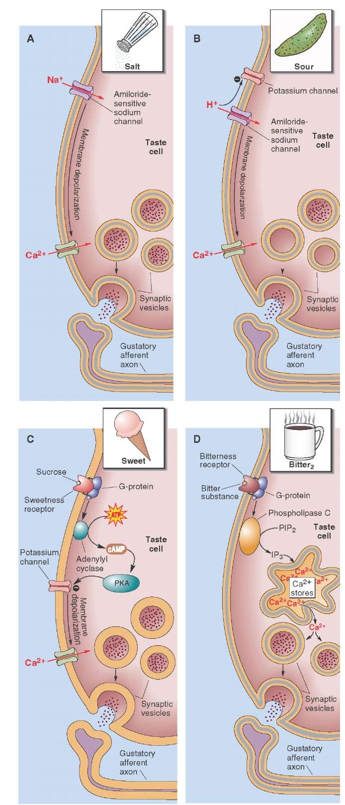
FIGURE 18-5 Transduction of taste stimulus. (A) The transduction of salt taste is mediated by the influx of Na+ (sodium) through the amiloride-sensitive Na+ channels. (B) Sour taste is mediated by closure of voltage-dependent K+ (potassium) channels. (C) Sweet taste is mediated by activation of G proteins and adenylate cyclase, which increases the levels of cyclic adenosine monophosphate (cAMP). (D) Bitter substances activate a G protein that activates phospholipase C and generates inositol triphosphate (IP3). Ca2+ = calcium; ATP = adenosine triphosphate; PKA = protein kinase A; PIP2 = phosphatidylinositol 4,5-biphosphate.
This initial step of depolarization is brought about by opening or closing of different channels. For example, the transduction of salty taste is mediated by generation of a receptor potential due to influx of Na+ through the amiloride-sensitive Na+ channels (Fig. 18-5A). Sour taste, elicited by acids, is mediated by depolarization of the receptor cell due to closure of voltage-dependent K+ channels (Fig. 18-5B). Other mechanisms for mediation of taste sensation involve activation of a G protein that, in turn, activates a cascade of events resulting in transmitter release.For example, substances that generate the sense of sweet flavor (e.g., sugars) act on receptors that are coupled with Gs proteins. Activation of G proteins results in activation of adenylate cyclase (ade-nylyl cyclase), which increases the levels of cAMP. cAMP activates a phosphokinase that depolarizes the receptor cells by closing K+ channels (Fig. 18-5C). Bitter substances activate a G protein, which, in turn, activates phospholi-pase C and generates the IP3 second-messenger system. IP3 releases Ca2+ from intracellular stores (Fig. 18-5D). Activation of different second-messenger systems eventually causes opening of voltage-dependent Ca2+ channels and influx of Ca2+, which results in transmitter release and activation of afferent nerve terminals at the base of the receptor cell.
Central Pathways
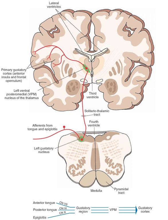
The taste buds on the anterior two thirds of the tongue are innervated by the facial nerve (CN VII), taste buds on the posterior one third of the tongue are innervated by the glossopharyngeal nerve (CN IX), and the taste buds on the epiglottis and pharyngeal walls are innervated by the vagus nerve (CN X) (Fig. 18-6). The afferent terminals of the facial nerve carry sweet, sour, and salty sensations; whereas those of the glossopharyngeal nerve carry sour and bitter sensations.
Pseudo-unipolar neurons mediate the sensation of taste. The pseudo-unipolar neurons mediating the sensation of taste via the facial nerve (CN VII) are located in the geniculate ganglion, which is situated in the petrous portion of the temporal bone. The peripheral processes of these neurons travel in the facial nerve, which exits the cranium at the stylomastoid foramen. At this level, the peripheral processes of the sensory neurons exit from the facial nerve and form the chorda tympani nerve, which crosses the cavity of the middle ear (horizontally along the inner surface of the tympanum and over the manubrium of the malleus ossicle). The chorda tympani finally joins the lingual branch of the trigeminal nerve and innervates the taste buds on the anterior two thirds of the tongue. The central processes of sensory neurons in the geniculate ganglion travel in the intermediate nerve (adjacent to the facial nerve), enter the solitary tract, and terminate in the rostral portion (gustatory region) of the solitary nucleus (Fig. 18-6).
The unipolar neurons mediating the sensation of taste via the glossopharyngeal nerve (CN IX) are located in the inferior (petrosal) ganglion, which is located in the jugular foramen. The peripheral processes of these neurons travel in the glossopharyngeal nerve and finally innervate the taste buds on the posterior one third of the tongue. The central processes of sensory neurons in the petrosal ganglion travel in the glossopharyngeal nerve, enter the solitary tract, and also terminate in the rostral portion of the solitary nucleus, which is known as the gustatory nucleus (Fig. 18-6).
FIGURE 18-6 Central pathways that mediate taste sensation. The unipolar neurons mediating the sensation of taste via the facial, glossopharyngeal, and vagus nerves are located in the geniculate, inferior (petrosal), and inferior (nodose) ganglia, respectively. The peripheral processes of these neurons terminate in the rostral portion (gustatory region) of the solitary nucleus. The axons of secondary neurons located in the solitary nucleus ascend in the solitario-thalamic tract and terminate in the ventral posteromedial nucleus (VPM) of the thalamus. The neurons in VPM send their projections to the taste (gustatory) area located between the anterior insula and the frontal operculum in the ipsilateral cerebral cortex. CN = cranial nerve.
The unipolar neurons mediating the sensation of taste via the vagus nerve (CN X) are located in the inferior (nodose) ganglion, which is located just below the jugular foramen. The peripheral processes of these neurons travel in the vagus nerve and innervate taste buds on the epiglottis. The central processes of the sensory neurons in the nodose ganglion travel in the vagus nerve, enter the solitary tract, and terminate in the gustatory nucleus (Fig. 18-6).
The axons of secondary neurons located in the gustatory nucleus ascend in the solitario-thalamic tract (near the medial lemniscus) and terminate in the ventral posteromedial nucleus (VPM) of the thalamus (Fig 18-6). The neurons in the VPM send their projections to the taste (gustatory) area located between the anterior insula and the frontal operculum in the ipsilateral cerebral cortex. Other fibers of the taste system pass from the solitary nucleus to the amygdala, either directly or indirectly, via connections in the pedunculopontine nucleus (i.e., pontine taste nucleus) of the reticular formation.
Taste Perception
Our understanding of the neural basis of taste perception is incomplete. Two theories have been proposed to explain the perception of taste. One theory, the specific pathway theory, states that individual taste receptors respond to a single taste stimulus (e.g., sweet taste). This information is transmitted to specific populations of neurons within the taste pathway for perception of that taste quality. The second theory is based on the concept of an across-fiber pattern coding. This theory states that individual taste receptors respond to more than one modality of taste. Therefore, the perception of a given taste may involve processing of the response patterns of many taste afferents by cortical neurons within the cortical taste area. Such a response gives us the ability to discern nuances of flavor, as in wine or other fine foods.
Clinical Conditions in Which the Taste Sensation is Altered
Ageusia is loss of taste sensation. Damage to the nerves innervating the taste buds may cause total ageusia (loss of all taste sensation), partial ageusia (loss of a particular taste sensation), or hypogeusia (decreased sensation of taste) depending on the extent of damage.
Clinical Case
History
Celia is a 32-year-old woman who had no medical problems. One day while shopping in a department store,she was admiring a dress while walking and did not notice a clothing rack rapidly being pushed in her direction. Because she was not paying attention to what was in front of her, she collided with the clothing rack, hitting her face. She immediately noticed pain in her nose and forehead. Because her nose was bleeding, she was taken to the emergency room, where skull x-rays were performed, revealing a small fracture in the cribriform plate that was too small for any therapy. She was sent home with pain medication and told to return if there were any further sequelae.
Examination
Two weeks later, after the swelling and bleeding had subsided somewhat, while eating dinner at a restaurant, Celia noted that she was unable to smell the food.
This continued with subsequent meals, so she consulted a neurologist who tested her sense of smell with several substances, including coffee grounds. He concluded that her anosmia (inability to smell) was a result of her head trauma and appeared to exist on both sides of her nose.
Explanation
Celia has anosmia resulting from trauma to the cribriform plate. This causes damage to the filaments of the receptor cells for smell as they pass through the cribriform plate. Depending on the extent of the injury, the sense of smell might return in approximately one third of cases in a period of days to weeks.
SUMMARY TABLE
Pathways That Mediate Olfactory and Taste Sensations
|
Receptors |
Sensory Neuron |
Pathways |
|
|
Central Olfactory Pathways |
|
|
Olfactory knob (expanded dendrite of an olfactory cell) located on the surface of olfactory mucosa; 10-20 cilia arising from the olfactory knob spread on the surface of the olfactory mucosa and are involved in sensory transduction of smell |
Sensory neuron located in the olfactory mucosa of the nasal cavity below the cribiform plate (rather than in a ganglion as in other sensory systems) |
1. Axons arising from sensory neurons form small clusters that are collectively called olfactory nerve |
|
2. Axons of olfactory nerve project to ipsilateral olfactory bulb |
||
|
3. Olfactory bulb contains different layers (olfactory nerve layer,glomerular layer,external plexiform layer, mitral cell layer, and inner plexiform layer) |
||
|
4. Largest bundle of fibers from mitral and tufted cells exits from olfactory bulb in lateral olfactory tract and projects to primary olfactory cortex (piriform cortex), amygdala, and entorhinal cortex |
||
|
5. Some fibers from mitral and tufted cells exit olfactory tract via medial olfactory tract |
||
|
6. Note that olfactory projection differs from other sensory systems in that projection reaches prefrontal cortex without making a synapse in thalamus; in particular, neurons in amygadala project directly to prefrontal cortex |
||
|
7. Some neurons in the piriform cortex project to medio-dorsal thalamic nucleus, which then sends fibers to wide areas of frontal lobe, including prefrontal cortex |
||
|
8. Neurons in entorhinal cortex project to the hippocampus via the perforant pathway |
||
|
9. Some fibers from mitral and tufted cells exit via medial olfactory tract, which also contains fibers arising from contralateral anterior olfactory nucleus; axons in medial olfactory tract project ipsilaterally to basal limbic forebrain structures (substantia innominata, medial septal nucleus, and bed nucleus of stria terminalis) |
||
|
10. It is believed that different glomeruli located in spatially distinct parts of olfactory bulb respond to specific odor-ants; olfactory signals become topographically organized within olfactory bulb; this arrangement provides a basis by which neuronal pools within prefrontal cortex can receive and transform olfactory signals into a conscious awareness of a specific odorant |
||
|
Receptors |
Sensory Neuron |
Pathways |
|
|
Central Olfactory Pathways |
|
|
11. Affective and emotional aspects of olfactory sensation are mediated by olfactory projections to limbic system (entorhinal cortex, hippocampal formation, medial septal nuclei, and amygdala); autonomic responses to olfactory stimuli are mediated via descending projections from the hypothalamus, midbrain PAG,and autonomic centers of lower brainstem and spinal cord |
||
|
Central Pathways That Mediate Taste Sensation |
||
|
Taste buds (papillae) located in protrusions on surface of tongue; each has a pore at its tip through which fluids containing chemical substances enter; taste bud contains taste receptor cells; afferent nerve terminals make contact with base of taste receptor cells |
Sensory neurons mediating taste sensation via CN VII,CN IX and CN X are located in geniculate, inferior (petrosal), and nodose ganglia |
1. Innervation of taste buds on anterior two thirds of tongue, posterior one third of tongue, epiglottis, and pharyngeal walls is provided by facial, glossopharyngeal, and vagus nerves, respectively |
|
2. Afferents in facial nerve carry sweet, salty, and sour sensations; those in glossopharyngeal nerve carry sour and bitter sensations; note that the concept that specific areas of tongue mediate specific taste sensations is not universally accepted, and it has been suggested that taste sensation arises from all regions of oral cavity |
||
|
3. Central processes of sensory neurons in geniculate, petrosal, and nodose ganglia travel in intermediate branch (adjacent to main trunk of facial nerve) and glossopharyngeal and vagus nerves, respectively, and terminate in rostral portion of solitary nucleus (gustatory region) |
||
|
4. Axons of secondary neurons located in gustatory region ascend in solitario-thalamic tract and terminate in VPM of thalamus; neurons in VPM project to taste (gustatory) area located between anterior insula and frontal operculum in ipsilateral cerebral cortex; other fibers from solitary nucleus project to amygdala, either directly or indirectly, via connections in pedunculopontine nucleus of reticular formation (i.e., pontine taste nucleus) |
||
|
5. Two theories of taste perception: (1) individual taste receptors respond to a single taste stimulus, and this information is transmitted to specific populations of neurons within taste pathway for perception of that taste quality, (2) individual taste receptors respond to more than one modality of taste, and response patterns of many taste afferents are processed by neurons within cortical taste area |
||
CN=cranial nerve; PAG = periaqueductal gray; VPN=ventral posteromedial nucleus.


