In the previous topic, we examined the organization of the medulla. In this topic, a parallel analysis will be made with respect to the pons and cerebellum. Similar to the medulla, the pons consists of different kinds of nuclear groups belonging to cranial nerves as well as other groups that are unrelated to cranial nerves. There are also long ascending and descending fiber pathways that pass through the pons, the majority of which are also present in the medulla. In addition, two of the cerebellar peduncles linking the cerebellum with the brainstem (the middle and superior cerebellar peduncles) pass directly from or through the pons, respectively.
Gross Anatomical View of the Pons
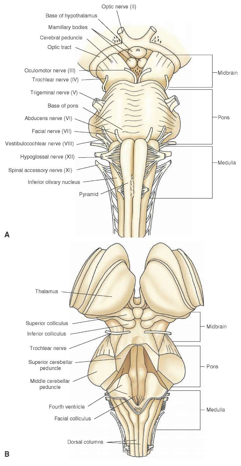
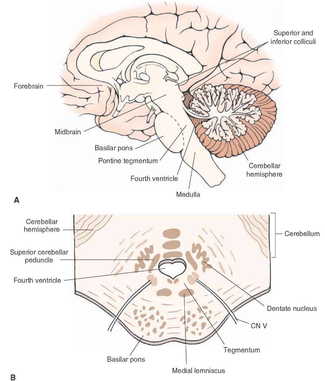
In its gross appearance, the pons serves as a connection between the medulla, which is attached to its caudal end, and the midbrain, which is attached at its rostral end (Fig. 11-1). From an anatomical perspective, the pons is typically divided into two regions, a ventral part, called the basilar pons, and a dorsal part, called the tegmentum (Fig. 11-2). The basilar pons consists of two different groups of fibers and neurons distributed throughout much of this region. One group of fibers, present throughout both the dorsoventral and rostro-caudal aspects of the basilar pons, runs in a transverse direction across the pons. This massive group of fibers is, thus, referred to as transverse pontine fibers (pontocerebellar) and arises from the neurons scattered throughout the basilar pons called deep pontine nuclei. The second group of fibers consists of cor-ticospinal and corticobulbar fibers, which descend from the cerebral cortex and pass in a longitudinal manner with respect to the neuraxis of the brainstem to the spinal cord and lower brainstem, respectively, and are, thus, positioned perpendicular to the orientation of the transverse pontine fibers.
FIGURE 11-1 Ventral and dorsal views of the brainstem. (A) View of the ventral surface of the brainstem illustrating the position of the pons in relation to the medulla and midbrain and the loci of the cranial nerves (CN). Note that CN VI and VII exit medially and laterally, respectively, at the level of the caudal pons, and CN V exits laterally at the level of the middle of the pons. (B) View of the dorsal surface of the brainstem looking down into the fourth ventricle, illustrating the positions of the pons in relation to the medulla and midbrain. The cerebellum was removed to demonstrate the position occupied by the fourth ventricle. Note the positions occupied by the superior and middle cerebellar peduncles.
Along the midline of the basilar pons is a groove called the basilar sulcus, which is formed by the basilar artery. Laterally, another massive fiber bundle, called the middle cerebellar peduncle, is a continuation of the transverse pontine fibers. This peduncle passes laterally and dorsally into the cerebellum from the lateral aspect of the pons, thus forming a "bridge" between these two structures. The dorsal aspect of the pons (tegmentum) forms the floor of the fourth ventricle and the roof of the pons. Along the floor of the fourth ventricle, a bulge can be identified cau-dally along its medial aspect. This is called the facial colliculus and is formed by the presence of the fibers of the facial nerve that pass over the dorsal aspect of the abdu-cens nucleus (Fig. 11-1B). The lateral walls of the fourth ventricle are formed by the superior cerebellar peduncles that arise mainly from the cerebellum and enter the brainstem from a dorsolateral position at the level of the rostral pons.
FIGURE 11-2 Sagittal view of the brainstem. (A) Midsagittal section through the neuraxis of the brain showing the relationship of the pons to the medulla, midbrain, cerebellum, and forebrain. Note that the pons can be divided into two regions: a ventral aspect, the basilar pons, and a dorsal aspect, the tegmentum. (B) A cross section taken through the middle of the pons reveals the division of the pons into the basilar and tegmental regions. CN = cranial nerve.\
The tegmentum of the pons also consists of nuclei and fiber pathways. Nuclear groups include groups associated with cranial nerves as well as those that are unrelated to cranial nerves but that serve important sensory, motor, autonomic, and behavioral functions. The fiber pathways in this region consist of axons that may arise in the pons and that run either dorsally or caudally. In addition, this region also contains other ascending or descending fiber pathways, which originate in the brainstem, spinal cord, other parts of the brainstem, or forebrain. Details concerning the nature of these cell groups and fiber pathways are considered in the following sections.
Internal Organization of the Pons
Fiber Pathways
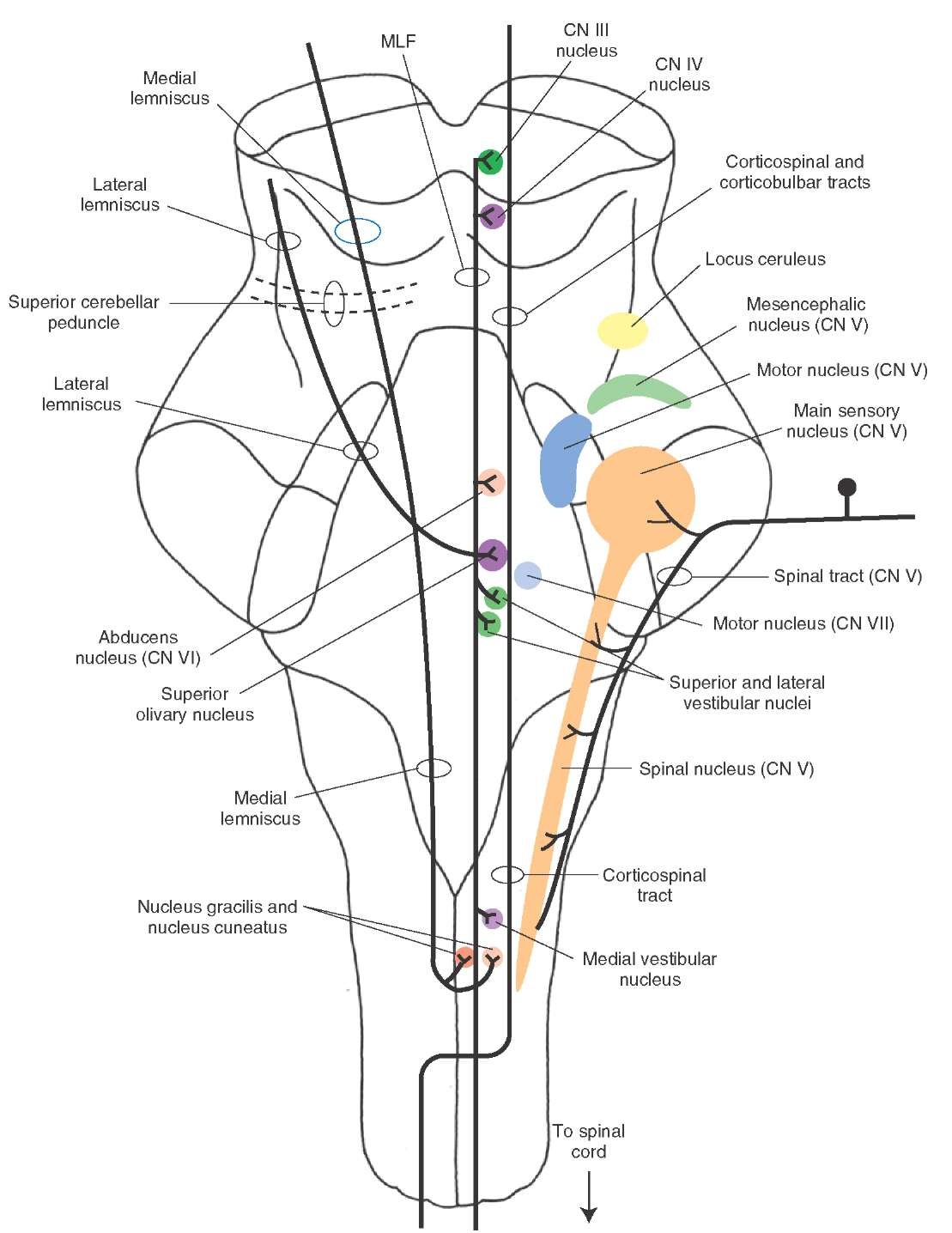
Many of the same pathways present within the medulla are also present within the pons. Two of the most prominent ascending tracts include the medial lemniscus and medial longitudinal fasciculus (MLF) (Fig. 11-3). Note that, as the medial lemniscus passes in a rostral direction within the pontine tegmentum, it becomes oriented more laterally and somewhat more dorsally until it reaches its most lateral and dorsal position in the caudal diencephalon where it synapses in the ventral posterolateral nucleus of the thalamus.In contrast, the MLF retains its dorsomedial position as it ascends towards the midbrain. In addition, other pathways are situated in a lateral position within the pontine tegmentum as they ascend to the midbrain and more rostral levels. These pathways include the spinothalamic and trigeminothalamic pathways that mediate somatosensory information from the body and head region to the ventral posterolateral and ventral posteromedial nuclei of the thalamus, respectively. These two pathways are sandwiched between the lateral lemniscus, a part of the auditory relay circuit, and the medial lemniscus.
FIGURE 11-3 The general positions of the major nuclear groups and most prominent ascending and descending pathways that traverse the pons. CN = cranial nerve.
The major descending pathways include the corticobul-bar and corticospinal tracts (Fig. 11-3). These pathways maintain a position within the basilar aspect of the pons. Other pathways (not clearly discernible from normal sections of the pons) include the rubrospinal and tectospinal tracts. The rubrospinal tract occupies a position within the pons just dorsal to the medial lemniscus. The position of the tectospinal tract within the level of the pons is similar to its position within the medulla; namely, it lies dorsome-dially just ventral to the MLF. In addition, the dorsolateral aspect of the tegmentum of the pons (as well as the teg-mentum of the midbrain and medulla) contains descending fibers from the limbic system and hypothalamus that project to regions of the lower brainstem that mediate autonomic functions.


