abstract
The revolution of technology has lead to a change; from the analogic to the digital function of medical devices. Some of them were produced in the last years to improve the quality of images. Although the procedure of acquiring and using the devices has been very complicated, the analysis of the images is so dependable that a big amount of the annual budget is spent for their acquisition.
Introduction
The rapid development of science and the continuous manufacture of pioneer medical technological products, together with the modern requirements for high-quality medical services, led to the growth and introduction of modern technologies in the health sector. This development has been really impressive and rapid.
At the beginning of the 20th century, the progress of applied sciences, such as chemistry, physics, microbiology, physiology, pharmacology, and so forth enforced medical research, which resulted in the continuous discoveries in medical technology. In 1895, W. K. Roentgen discovered X-rays, which was a turning point for medical imaging and diagnostics in general. In the 1950s, there was the development of computerized systems, while in the 1960s, there were applications such as the transport of biological signals from equipped space missions and teletransfers of information.
To be more specific, the dynamic entry and the enforced intervention of sciences such as informatics gave an enormous impulse to the medical field and created new data for treatment and diagnosis. Thus, we now have the interaction and participation of many different scientific sectors, which aim at the best medical care. One of these sectors is the imaging and treatment of medical pictures, whose development and use are a crucial point in modern therapeutics.
Medical imaging is related to issues such as the descriptive principles of medical images and their elaboration, together with whatever they include. Therefore, it is easy for someone to understand the importance and the role of imaging in diagnosis, treatment, and recovery in general.
This topic will discuss the basic concepts, such as the analysis of medical images, their elaboration, various uses and applications, as well as an analysis of the functional requirements of such applications in order for them to be fulfilled.
DIGITAL ELABORATION OF IMAGES
The digital elaboration of images is actually a new sector of informatics and has been applied for only 15 years. Its development is revolutionary in the health field and constitutes a major contribution for the promotion of health.
The vast amount of optical information and the need for elaborating them led scientists and technicians to the discovery of storage means for the images and their elaboration by using computers combined with the development and improvement of biomedical equipment.
Digital elaboration, as the title itself declares, deals with the digital registration of images and their elaboration by computers. The objective of elaboration can be the quality improvement of images, the straining of recording or transmission noises, the compression of the amount of information, the storage of images, and their digital transmission and depiction.
In the picture below, we schematically represent all the necessary equipment that is needed to fulfill a digital registration of images to be elaborated by the appropriate personnel in order to give all the necessary information and conclusions.
Thus, everyone can understand that for the elaboration of three-dimensional medical images, a device for the admission of images is required, and this can be an axial tomographer, a magnetic tomographer, an ultrasound device, or even more developed depiction systems. A computer is also required, with all the necessary equipment in order to elaborate the images, analyse them, and store and transmit them, and finally all the appropriate exit devices that will project the images will be needed, which can be terminal stations, special films, and printers. Finally, it is very important that the whole system will support network communications so that all information can be available to the scientific community fast and safely with both quality and reliability. Before we introduce some of the depiction devices of three-dimensional medical images that are widely used by the scientific community nowadays, it is important to study some necessary notions for the comprehension of the way this elaboration of three-dimensional images occurs.
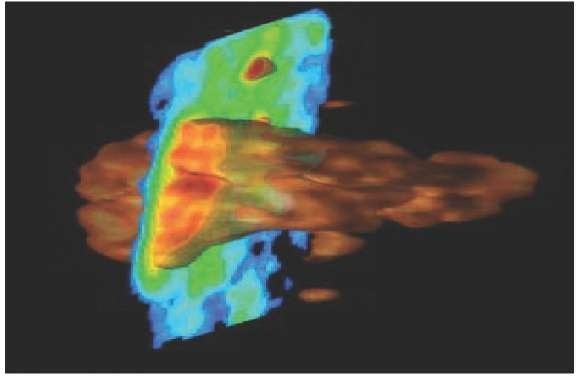
Figure 1. Three dimensional imaging
An image is a two-dimensional signal. Thus, for the analysis and elaboration of this signal, all the techniques and mathematic relations of digital signal elaboration can be used. Specifically, in the health field, we introduce the notion of modality, which is a biomedical signal that represents one view or one function of the organization. Therefore, the depiction devices receive modalities and with the appropriate processes turn them into images. The dimension of a signal depends on the number of independent variables it has. Consequently, the two-dimensional signals have as independent variables the two dimensions of the surface.
Therefore, the elements that should be included in a digital system for the elaboration of images are the following:
• Image Elaborator: This is the hardcore of the system that receives the image, temporarily stores and elaborates it on a low level, and finally demonstrates it on a primary level.
• Digitizer: It arithmetically represents the image in order for it to be received by a computer.
• Digital Computer: It performs an extensive elaboration.
• Storage Devices
• Screens and Recording Devices
We will now proceed to analyse the above elements in order to realize their participation in the elaboration of images, and at the same time we will present all the processes that occur at each level.
Image Elaborator
The evolution of technology, as mentioned above, was the vaulting bar for the development and evolution of medical depiction devices and machines, something that proved to be beneficial for health services. Today, there is a large number of medical apparatuses that fills any kind of diagnostic demands. More specifically, in the three-dimensional depiction area, there are now many options that suit every demand. The categories of medical images are as follows.
Figure 2. Image elaborator
• Digital abstraction angiography
• Ultrasound system
• Computed tomography (CT)
• Magnetic resonance imaging (MRI)
• Gamma camera
• Computed tomographies of nuclear medicine (single-photon-emission computed tomography [SPECT] and positron-emission tomography [PET])
Digital Abstraction Angiography
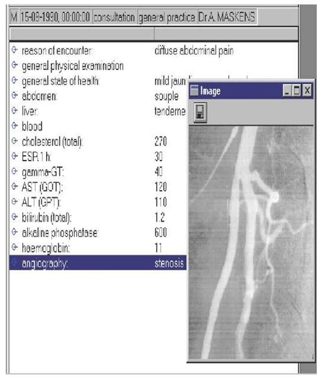
It is a medical diagnostic examination that depicts the condition of the vessels. It is extremely useful in the sector of cardiology and helps to with the prevention of many diseases of the cardiac system. Its characteristic is that it produces a vast number of data, especially as we move from one-dimensional systems to two-dimensional ones and from a still image to a moving one (video).
Ultrasound System
It depicts the resonance of high-frequency sound waves, which depend on the auditory properties of tissues that are produced by different organs and are examined as brightness in the image. One application of the ultrasound system is the ultrasound of the heart and vessels (Doppler), which is developed in real time and depicts the flux speed of the blood.
Computed Tomography
It uses X-rays for the creation of images, which means that the patient is exposed to radiation during the examination. The part of the body that needs to be examined receives X-rays through different transmission angles. With the use of mathematic transformations and calculations on the counted prices from these different angles, we receive images that are cross and plane sections. Each ray penetrating the body is recording densities of tissues.
Figure 3. Digital angiography
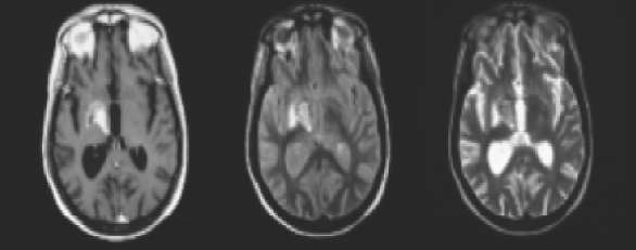
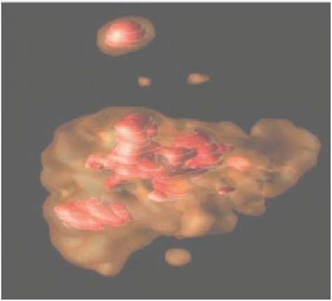
Figure 4. MRI depiction
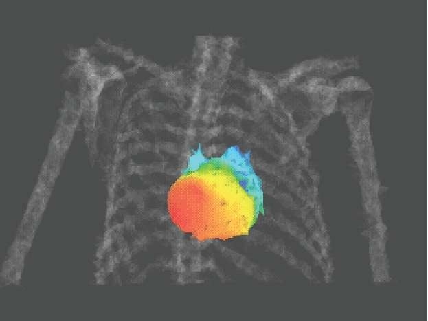
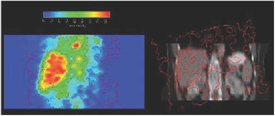
Figure 5. Depiction in SPECT
Magnetic Tomography
It is a nonpenetration diagnostic method that produces a series of images that represent biological differences among tissues. Furthermore, the patient does not have to be exposed to harmful radiation. It is based on the application system of a magnetic field in order to obtain magnetic tomographies.
By applying a magnetic field, the cells orient to the rotation frequency and thus we have the production of resonance through the pulse of radiofrequency. Afterward, we have a rest so that the cells can reorient to their initial positions. The produced signal depends on the density and type of cells and, thus, magnetic tomographies show a high contrast on the soft tissues.
Gamma camera
It is a diagnostic method that belongs to the field of nuclear medicine. It is based on the calculation of the position and concentration of a radioactive isotope that is provided to the patient before the examination. It gathers clinical information about the normal function while its discernibility is very low.
Computed Tomographies of Nuclear Medicine (PET and SPECT)
This section is about diagnostic examinations that are related to the physiology of the organization and are the most modern in technological terms in the field of diagnostics. SPECT involves the computed tomography of photon transmission, something that shows that the signal-production process is based on photon transmission. PET involves tomography with positron transmission.
The diagnostic devices mentioned are the means of acquiring medical images in analogous or digital forms. The device used may have the ability to store digitally; otherwise, the digitali-zation occurs with the use of a digitizer before storage. Next we have the process of compressing images.
Digital Image compression
The term compression is mentioned in a number of technicalities and algorithms aiming to reduce the required memory for the representation and storage of digital images. The storage of a digitalized image leads to a squandering of the memory of the computing system. Thus, in the medical field, where the amount of image information is enormous, a major problem rises because of the amount of memory required for image storage. For this reason, faster compression and decompression algorithms for images have been searched for. Besides sparing memory, compression provides a reduction in the time and the width needed for transmission.
The techniques for the compression of digital images can be divided into two categories.
In medicine, where even the smallest detail in depiction can be of vital importance, we choose lossless techniques.
Compression is based on redundant information that is included in the images. The more redundancy there is, the more is the achieved compression. The whole process is called source coding, and it is based on a system that includes the following:
• Transformer: It transforms the initial image into a more appropriate one for compression.
• Quanter: It quants the transformed image either with graduation or with vector.
• Coder: A sequence of bits corresponds with each quant level. The code can be of a specific or variable length.
Compression with no losses does not use a quanter system because a quant always shows a loss of information.
The used patterns of image compression are as follows:
• G3, G4, GBIG—binary images
• JPEG—firm images
• H.261 MPEG1, MPEG2, MPEG4, MPEG7— movable images
After the compression process, we have the process of storage to the PACS system and recuperation from RIS.
Storage of Medical Images and Their Recuperation
Medical images, according to the diagnostic device used, have large sizes due to the need for high-quality analysis and clearness, thus demanding a large storage space. The recuperation time differs according to the kind of examination and the demanded number of images in each examination.
When the image reaches the final stage of elaboration, the goals are then the improvement of the quality of the given information with the use of the appropriate graphic-elaboration software (e.g., clearness, brightness, contrast), and also the exploitation of specific information with the use of special operations and transformations like division and recombining.
Quality Improvement of Images
The quality improvement of images is achieved with the use of software that helps to reach the desired levels of analysis aiming to exploit and evaluate the information given from an image. During the process of recording images, certain deformities appear. These are:
• Dimness,
• Noise of recording, and
• Geometrical deformities.
All these deformities should be corrected in order to eliminate all kinds of alterations. The elimination of dimness is done with the process of restoration. The process of restoration is extremely important in the elaboration of moving images because movement creates dimness. In most cases, a filtering of the image is also required in order to remove sound. This can be achieved with various linear or nonlinear filters. Usually, nonlinear filters are preferred because they preserve the contrast of outlines, which are an important factor for human vision. Moreover, the general contrast of the image can be improved with special nonlinear techniques.
One other important procedure for image analysis is the recognition of outlines. Usually, the areas of an image are coloured with false colours.
Finally, we achieve improvement of quality by adjusting the proper clearness, brightness, and contrast with the use of appropriate technical equipment that allows us to exploit all these tools provided by the graphic software for quality improvement.
Image Division
By dividing a medical image, we are able to divide the image in different sections that show some cognitive interest according to the speciality under examination. Thus, according to the reason why the diagnostic examination occurs, the scientist can focus on a specific part of the image to process suitably and find the needed information without the redundancy of information to trouble him or her.
Furthermore, with the appropriate software, we can store to the computed system only those parts of the image that interest us, thus sparing storage space and making it more easy to recuperate the images from the used medical databases.
Medical Image Recombination
With medical image recombination, we can have a combination of different views from various depiction examinations (e.g., ultrasound+CT, MRI+PET, MRI+SPECT), giving us a three-dimensional representation of composition.
Indeed, medical image recombination is extremely important in medical depiction. Each diagnostic examination allows the scientist to extract specific information through it. With this recombination method, the scientist can have the elements and information provided by different diagnostic examinations in only one image, can elaborate them and analyse them in any way he or she desires, and, finally, can reproduce them in a three-dimensional environment. The whole undertaking occurs through complex graphic software, which should have certain characteristics in order to be an actual diagnostic tool in the hands of the scientist vs. an obstacle in his or her effort to do the job.
Figure 6. Division of image received by an MRI
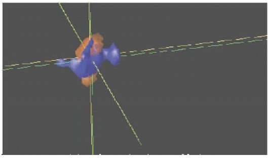
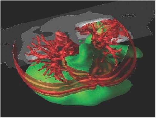
Figure 7. Image recombination MRI + PET + CT
Figure 8. Image recombination SPECT + CT
The characteristics of the elaboration software include the following:
• Friendly user interface
• Large number of choices that are easy to overcome
• Support of the established models of compression and image management
• Support of future applications
• Safe use
• Is easy to learn
conclusion and perspectives
Three-dimensional image elaboration is a very strong tool in medical diagnostics. Many of the diseases of tissue and microscopic interest are able to be diagnosed early and therefore can be cured. Especially benefited are the fields of research in medicine, pharmacology, and genetics, where the elaboration and depiction of three-dimensional models is extremely crucial for their development. The appearance of more and more evolved depiction devices and the growth of more powerful and more complete software for digital graphic elaboration will predispose even bigger steps of evolvement in the near future.
