Tracheal System
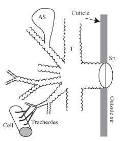
Insects have a tracheal respiratory system in which oxygen and carbon dioxide travel primarily through air-filled tubes called tracheae. Usually the tracheal system penetrates the cuticle via closeable valves called spiracles and ends near or within the tissues in tiny tubes called tracheoles (Fig. 1). The tracheae primarily serve as pipes that transport gases between the spiracles and the trache-oles, whereas the thin-walled tracheoles are thought to be the main sites of gas exchange with the tissues. However, in many insects, the tracheae are compressible, and dilations of the tracheae form thin-walled air sacs that together serve as bellows for enhancing the flow of gases through the tracheal system. The organization of the tra-cheal system varies dramatically among insects, with spiracle number ranging from 0 to 20 and with tracheal branching patterns varying widely across species, between body regions, and during the developmental stages of holometabolous insects.
SPIRACLES
Spiracles are the openings of the tracheal system on the integument of the insect. Some apterygote and larval insects lack valves in their spiracles and therefore have trachea that are always open to the environment, although these are often covered with sieve-like plates. However, most spiracles have valve-like structures that allow them to close (Fig. 2). Spiracles range tremendously from 27 fim2 in area for the second spiracle of the giant Petrognatha beetle to only a few microns

FIGURE 1 An overview of the tracheal system. Spiracles (Sp) in the integument connect to tracheae (T), which branch repeatedly within the insect, eventually leading to the tracheoles, which are located near most cells. In some insects, soft, collapsible air sacs (AS) occur.
square. Often the outside of the spiracle is fringed with hairs or covered with a filter to help resist entry of dust, water, or parasites.
The valve that closes the spiracle takes various forms but can appear as two lips of cuticle or as a bar of cuticle that pinches the trachea shut (Fig. 2). The movement of the valve usually depends on the action of muscles that insert on or near it, and elastic elements in the cuticle. Some thoracic spiracles have only a closer muscle, and elastic elements tend to open the spiracular valve (Fig. 2A). Often the valve is recessed behind the atrium, an enclosed space behind the external opening (Fig. 2B). The most common situation (true for all abdominal spiracles studied to date) is for the spiracle to be recessed behind an atrium and to have both opener and closer muscles (Fig. 2B).
TRACHEAE
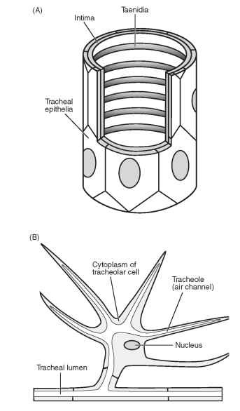
Tracheae, the largest tubes of the tracheal system, range in diameter from a few millimeters to about 1 um. The tracheal wall (Fig. 3A) contains an epithelial cell layer that secretes a basement membrane that forms the outermost layer of the tracheal wall. Internally, the tra-cheal cells secrete the intima, which contains protein and chitin fibers. The intima forms spiral folds known as taenidia. In general, it is thought that the taenidia prevent the trachea from collapsing inward, much like the rings of a vacuum hose, yet allow the trachea to flex. However, in the tracheae of some insects, the taenidia are widely spaced within the tracheal wall, and the intima is quite flexible so that the tracheae are compressible. Because tracheal walls are relatively thick, oxygen is not believed to pass out of the larger trachea in appreciable amounts. Thus, the primary funcstion of the trachea is to transport gases between the tracheoles and the spiracles. However, because carbon dioxide can diffuse more easily through tissue than oxygen, it is possible that a significant portion of the carbon dioxide emitted may pass through the tracheal walls.
TRACHEOLES
Tracheoles, the small tubes that form the terminal endings of the tracheal system, range from 1 to 0.1 um in diameter. Tracheoles are formed within single tracheolar cells (Fig. 3B). These tracheolar cells
![Spiracular structure and function. (A) Schematic side view of the locust metathoracic spiracle, a one-muscle spiracle with an external valve. [After Chapman, R.F. (1998). "The Insects: Structure and Function," Fig. 17.11c. Cambridge University Press, Cambridge, U.K., with permission of the publisher.] When the closer muscle (CM) contracts, the lips of the valve come together. When the closer muscle relaxes, ligaments (L) attached to the cuticle passively pull the valve lips apart. (B) Schematic view from the inside of the animal of the abdominal spiracle of a butterfly, a two-muscle spiracle with an internal valve.When the opener muscle (OM) contracts, it pulls on a lever which pulls the bar away from the bow, opening the atrium (A). When the closer muscle (CM) contracts, it pulls the bar against the bow, collapsing the walls of the atrium together. Spiracular structure and function. (A) Schematic side view of the locust metathoracic spiracle, a one-muscle spiracle with an external valve. [After Chapman, R.F. (1998). "The Insects: Structure and Function," Fig. 17.11c. Cambridge University Press, Cambridge, U.K., with permission of the publisher.] When the closer muscle (CM) contracts, the lips of the valve come together. When the closer muscle relaxes, ligaments (L) attached to the cuticle passively pull the valve lips apart. (B) Schematic view from the inside of the animal of the abdominal spiracle of a butterfly, a two-muscle spiracle with an internal valve.When the opener muscle (OM) contracts, it pulls on a lever which pulls the bar away from the bow, opening the atrium (A). When the closer muscle (CM) contracts, it pulls the bar against the bow, collapsing the walls of the atrium together.](http://lh3.ggpht.com/_X6JnoL0U4BY/S8H7y04OagI/AAAAAAAAYzo/tXRXYKqMC9U/tmp5B30_thumb.jpg?imgmax=800)
FIGURE 2 Spiracular structure and function. (A) Schematic side view of the locust metathoracic spiracle, a one-muscle spiracle with an external valve. [After Chapman, R.F. (1998). "The Insects: Structure and Function," Fig. 17.11c. Cambridge University Press, Cambridge, U.K., with permission of the publisher.] When the closer muscle (CM) contracts, the lips of the valve come together. When the closer muscle relaxes, ligaments (L) attached to the cuticle passively pull the valve lips apart. (B) Schematic view from the inside of the animal of the abdominal spiracle of a butterfly, a two-muscle spiracle with an internal valve.When the opener muscle (OM) contracts, it pulls on a lever which pulls the bar away from the bow, opening the atrium (A). When the closer muscle (CM) contracts, it pulls the bar against the bow, collapsing the walls of the atrium together.
have many branching processes, some of which contain an air-filled channel (the tracheole) that connects to the air-filled lumen of the trachea (Fig. 3B). Tracheole walls are capable of transporting oxygen at high rates by diffusion because they are thin (usually <0.1 um) and have a very large surface area to volume ratio. Thus, the tracheoles are likely the major site of gas exchange between the tissues and the tra-cheal system.
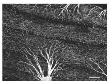
Tracheoles are particularly dense in metabolically active tissues such as flight muscle (Fig. 4). Most tracheoles occur outside of the cells of the insect’s body, but sometimes in histological sections they appear to be within cells, particularly in flight muscle. These tracheoles are believed to enter the flight muscle via infoldings of the muscle plasma membrane, and so they are actually extracellular. Most tracheaoles extend parallel to the muscle fibers. The high tracheolar densities and penetration of flight muscle cells by tracheoles allow flying insects to achieve oxygen consumption rates that are among the highest in the animal kingdom.
AIR SACS, AERIFEROUS TRACHEA,
AND TRACHEAL LUNGS
In active insects, particularly those with a rigid cuticle, portions of the tracheal system are enlarged to form thin-walled air sacs. These

FIGURE 3 (A) Structure of a trachea. The basement membrane which encircles the tracheal epithelia has been omitted. (B) A tra-cheolar cell connected to a trachea.
air sacs lack taenidia and, so, collapse and expand with variation in hemolymph pressure. Air sacs are common in adult flying insects of many orders including Diptera, Hymenoptera, Lepidoptera, Odonata, and Orthoptera. These air sacs serve as bellows to enhance the movement of air through the tracheal system. For insects that have them, air sacs often account for the majority of the volume of the tracheal system. Of insects that lack air sacs (such as cockroaches, tenebrionid or carabid beetles or Dictyoptera), many have compressible tracheae that may also function as bellows.
Tracheal lungs, or aeriferous tracheae, are similar to air sacs in that they are thin-walled expansions of the tracheal system with few taenidia that float freely in the hemolymph. Their primary function is believed to be oxygen delivery to tissues that lack tracheoles, such as hematocytes and some ovaries and Malpighian tubules. In this case,

FIGURE 4 Confocal image of tracheae branching into tracheoles supplying oxygen to the flight muscles of a fruit fly, Drosophila mela-nogaster Scale bar: 50 |im.
the oxygen must diffuse through the thin wall of the tracheal lung, through the hemolymph, and to the tissues. These tracheal lungs have been reported for lepidopteran larvae, which apparently lack hemocy-anin in the hemolymph; it would be interesting to look for such structures in the insects that utilize respiratory pigments in the blood.
DEVELOPMENT
The development of the tracheal system has been well studied in Drosophila melanogaster, and is serving as model for the development of the cardiovascular and respiratory systems of vertebrates. In flies, the tracheal system develops embryonically from epithelial sacs of only 80 cells per segment into its branching structure of tracheae and tracheoles without mitosis or cell death. Primary branching of the tracheae is induced by secretion of a local chemoattractant termed fibroblast growth factor (the gene is named Branchless) secreted by local tissues. The primary branches are formed of 3-20 cells organized into tubes. Some cells become terminal cells and ramify in response to fibroblast growth factor to become tracheae, with densities of tracheoles modulated by local oxygen levels.
In general, the size of the tracheal system increases with age in order to support the increased gas exchange needs of the larger insect. During the molts, the cuticular lining of the trachea is drawn out of the spiracle with the old integument. Major changes in tra-cheal structure, including changes in spiracle number and tracheal system organization, can occur at each molt and during the pupal period for endopterygote insects. In Drosophila, the adult tracheal system is formed from both imaginal disc cells and larval tracheal cells that activate and proliferate.
Changes in tracheal system structure are not limited to molting periods, because the tracheoles can change structure within an instar. In the event of injury or oxygen-deprivation, local tracheoles grow and increase in branching. If no undamaged tracheoles are nearby, damaged tissues produce cytoplasmic threads that extend toward and attach to healthy tracheoles. These threads then contract, dragging the trache-ole and its respective trachea to the region of oxygen-deficient tissue.
Both tracheoles and trachea are fluid-filled in newly hatched insects, and fluid fills the space between the old and new trachea at each molt.
Usually, this fluid is replaced with gas shortly after hatching or molting. In some cases, the spiracles must be open to the air for gas-filling of the tracheae to occur, suggesting that gas-filling occurs as fluid is actively absorbed by the tracheal and tracheole epithelia, with air entering through the spiracle to replace the absorbed fluid. However, in aquatic insects that lack spiracles, the tracheae also become gas-filled, indicating that this gas can be generated by the tissues or hemolymph.
ORGANIZATION
The organization of the tracheal system varies substantially among insects, at least partially in correlation with species ecology. One obvious variant is spiracle number. Some aquatic insects lack spiracles, which prevents water entry to the tracheae. Many dipteran larvae have only one pair of spiracles on their abdominal tip, which they extend into the air as they feed head-down in water. In contrast, many terrestrial insects have a full 10 pairs of spiracles. Insects also vary in the number of air sacs, the location and size of tracheal branches, and the number of longitudinal trachea.
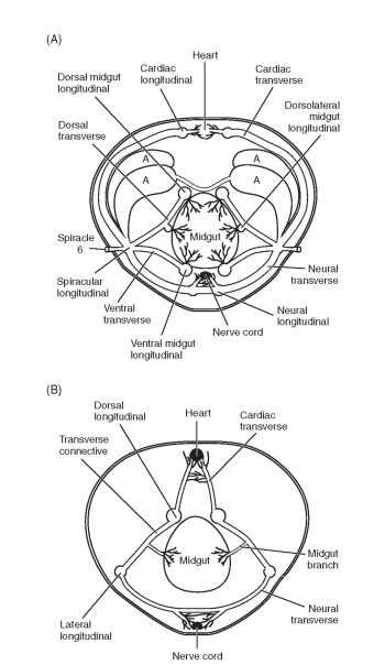
A glimpse of the cross-species variety can be gained by comparing the organization of the major tracheae in the sixth abdominal segment of an adult grasshopper and a larval fruit fly (Fig. 5). Grasshoppers have a spiracle on every segment, whereas fly larvae have only functional spiracles on the head and abdominal tip. In the grasshopper, transverse tracheae lead from the spiracles to longitudinal tracheae that run along the heart, ventral nerve cord, and several positions on the gut. Air sacs are prominent. In contrast, in flies, two large dorsal longitudinal trunks connect the two functional spiracles on each side, with branches extending from the dorsal trunk to the tissues and one other longitudinal trachea. Air sacs are absent.
Tracheal organization also varies with location within the insect. In most insects, the head segments lack spiracles, and so tracheae usually extend forward from the prothoracic spiracles to supply the tissues in the head; similar elaborations of the tracheae occur in the posterior direction in the 10th segment. Structures that extend away from the main body of the insect, such as legs, antennae, and wings, also lack spiracles and usually have long tracheae extending from the spiracle of the local body segment. Antennae, legs, and wings all are known to have accessory pulsatile structures that pump hemolymph and help circulate air through their long trachea. The tracheal organization in the thorax of flying insects is very different from that in the abdomen, as the thoraxes often contain extensive air sacs associated with the flight muscles.
Tracheal organization can also be strongly modified by developmental stage, especially in holometabolous insects. For example, many dipterans transform from aquatic larvae with one or two pairs of spiracles and no air sacs to adults with 10 pairs of spiracles and extensive air sacs, a transition that enables high oxygen delivery and flight. Clearly, flexibility in tracheal structure and function is a key trait in the success, adaptability, and diversity of insects.
As insects increase in size, it appears that they must invest proportionally more of their body in tracheal structures. This trend has been observed in growing grasshoppers and across species of beetles. It is not clear why this is true, but it may be as simple as the need for increased diffusing area to overcome the longer diffusion distances down the longer tracheae in bigger insects. At least in beetles, this trend is particularly true in the leg coxae, with larger beetles having proportionally more of the coxal lumen filled with tracheae. Extrapolation of this trend suggests that the largest extant beetles may begin to experience spatial limitations with the tracheae filling nearly 90% of the coxal lumen, suggesting that insect maximal body size may be limited by the need of larger insects for proportionally more tracheae.

FIGURE 5 (A) Cross section of the tracheal system at the sixth abdominal segment of an adult grasshopper (Schistocerca americana), an insect with air sacs and 10 paired spiracles. (B) Cross section of the tracheal system at the sixth abdominal segment of an embryonic or larval fruit fly (Drosophila melanogaster), an insect without air sacs and with only the first and tenth spiracles functional. In both insects, longitudinal tracheal trunks run the length of the animal, connecting the thoracic and abdominal segments, but longitudinal trunks are much more numerous in the grasshopper.
