Dental Arch Form
The teeth are positioned on the maxilla and mandible in such a way as to produce a curved arch when viewed from the occlusal surface (Figure 16-14). This arch form is in large part determined by the shape of the underlying basal bone.
Figure 16-14 The curvature of the maxillary (A) and mandibular (B) arches as seen from the occlusal (horizontal) plane tends to be maintained even though the tipped third molars alter the curvature (curve of Spee) of the arches as seen from the sagittal plane.
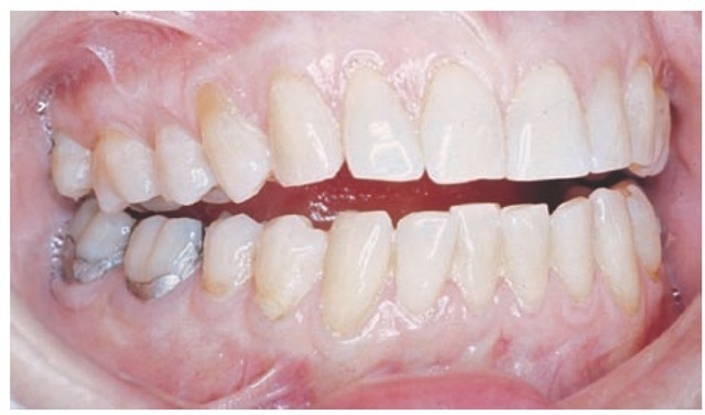
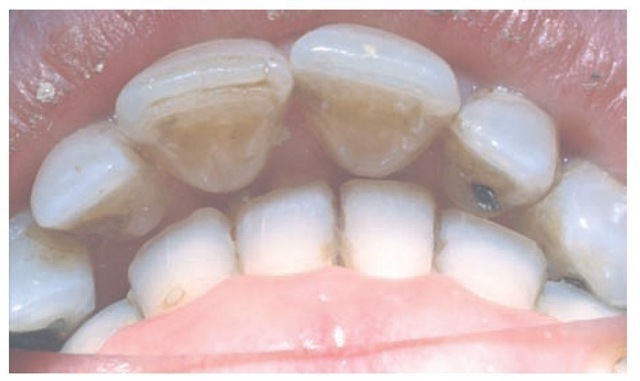
Malpositioning of individual teeth does not alter the arch form. When multiple teeth are misplaced, however, then irregularities or asymmetries may develop in arch form. A tapered arch form generally occurs in the maxillary arch and is quite often the result of a pathological narrowing of the anterior maxilla. Less commonly, a severe thumb-sucking habit may result in arch narrowing of the anterior maxilla (Figure 16-15).
The basic pattern of tooth position is the arch. On the basis of qualitative observations, anthropologists have described the general shape of the palatal arch as being paraboloid, U-shaped, ellipsoid, rotund, and horseshoe-shaped.29,30 An arch has long been known architecturally (as the word architecture itself implies) as a strong, stable arrangement with forces being transmitted normal to the apex of a catenary curve.31,32 The shape of the arch form of the facial surfaces of the teeth was thought by Currier33 to be a segment of an ellipse. In the past, interest in arch form was directed toward finding an "ideal" or basic mean arch form pattern that functionally interrelates alveolar bone and teeth and could have clinical application. However, any ideal arch pattern tends to ignore variance, a clinical reality which suggests that adaptation mechanisms are more important for occlusal stability than any ideal template.
Figure 16-15 Anterior open bite.
Figure 16-16 The terms overjet and overbite are commonly used to describe horizontal and vertical overlap of teeth.
Changes in arch form, within anatomical limits, do not have any significant effect on occlusion unless the change is in only one of the two dental arches. Discrepancies in arch form between the maxillary and mandibular arches generally result in poor occlusal relationships. Arch form distortion in only one arch can be advantageous when the basal bone structure is incorrectly positioned, as in severe mandibular retrognathism or prognathism. In such cases, the arch form distortion in one arch allows a better occlusion on the posterior aspect than is otherwise possible.
OVERLAP OF THE TEETH
The arch form of the maxilla tends to be larger than that of the mandible. As a result, the maxillary teeth "overhang" the mandibular teeth when the teeth are in centric occlusion (the position of maximal intercuspation). The lateral or anteroposterior aspect of this overhang is called overjet, a term that can be made more specific as indicated in Figure 16-16. This relationship of the arches and teeth has functional significance, including the possibility of increased duration of occlusal contacts in protrusive and lateral movements in incising and mastication.
The significance of vertical and horizontal overlap has to be related to mastication, jaw movements, speech, type of diet, and esthetics. Excessive vertical overlap of the anterior teeth may result in tissue impingement and is referred to as an impinging overbite (Figure 16-17). Correction is not simply a matter of trying to increase vertical dimension by restorations on posterior teeth. Orthodontics is generally required, and sometimes orthognathic surgery is recommended.
Gingivitis and periodontitis may occur from continued impinging overbite. The degree of vertical and horizontal overlap should be sufficient to allow jaw movement in function without interference. There should be sufficient vertical overlap (with the cuspid providing the primary guidance) to enable the disocclusion of the posterior teeth. Such movement in masticatory function is controlled by neuromuscular mechanisms developed out of past learning in relation to physical contact of the teeth. When protective reflexes are bypassed in parafunction, trauma from occlusion involving the teeth, supporting structures, and TMJs may occur. However, aside from cheek biting as a result of insufficient horizontal overlap of the molars and trauma to the gingiva from an impinging overbite, no convincing evidence shows that a certain degree of overbite or overjet is optimal for effective mastication or stability of the occlusion. Providing the correct vertical and/or horizontal overlap requires appropriate knowledge of dental morphology, esthetics, phonetics, restorative dentistry, function, and orthodontics.
The overlapping of the maxillary teeth over the mandibular teeth has a protective feature: during opening and closing movements of the jaws, the cheeks, lips, and tongue are less likely to be caught. Because the facial occlusal margins of the maxillary teeth extend beyond the facial occlusal margins of the mandibular teeth and the linguo-occlusal margins of the mandibular teeth extend lingually in relation to the linguo-occlusal margins of the maxillary teeth, the soft tissues are displaced during the act of closure until the teeth have had an opportunity to come together in occlusal contact. Cheek biting is commonly associated with dental restorations of second permanent molars that have been made with an end-to-end occlusal relationship (i.e., without overjet).
Figure 16-17 Impinging overbite.
CURVATURES OF OCCLUSAL PLANES
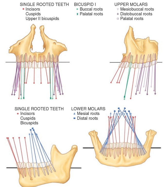
The occlusal surfaces of the dental arches do not generally conform to a flat plane (e.g., the mandibular arch has one or more curved planes conforming to the arrangement of the teeth in the dental arches). Perhaps the most well known is the curve of von Spee, who noted that the cusps and incisal ridges of the teeth tended to display a curved alignment when the arches were observed from a point opposite the first molars. This alignment (Figure 16-18) is spoken of as the compensating curve or curve of Spee.34 This curvature is within the sagittal plane only. Monson35 visualized a three-dimensional spherical curvature involving both the right and left bicuspid and molar cusps and the right and left condyles. It was supposed that the center of a sphere with an 8-inch diameter was the vector for converging lines of masticatory forces passing through the center of the teeth and that the occlusal surfaces of the molar teeth were congruent with the surface of a sphere of some dimension. This hypothesis was not supported by research by Dempster et al36 (i.e., the longitudinal axes of the roots of the teeth do not converge toward a common center) (Figure 16-19). Although the idea was incorporated into complete dentures and the design of some early articulators,37,38 such curvature has not been accepted as a goal of treatment even for dentures. However, the curvature of the occlusal plane such as the curve of Spee does have clinical significance in relation to tooth guid-ance—that is, canine and/or incisal guidance as applied in orthodontics and restorative dentistry. In these, disocclusion of the posterior teeth is desired during anterior (protrusive) movements.
Orthodontists tend to use no more than a slight arc of the curve of Spee when finalizing the occlusion, possibly to decrease the amount of vertical overlap of the maxillary canines and incisors needed to cause posterior disocclusion in protrusive movements and to decrease the frequency of relapse.
Figure 16-18 Curvature of the arch in the sagittal plane can be seen extending through the cusp tips from the third molar to the incisors.
Figure 16-19 Orientation of the crowns and roots. Top, Maxillary teeth in frontal (left) and lateral (right) view; bottom, mandibular teeth in lateral (left) and frontal (right) view.
Increasing the curve of Spee can compensate for smaller maxillary anterior teeth, especially lateral incisors. Reducing the curve of Spee can reduce the vertical overlap of the teeth. Any minimal standard, such as that the deepest curve should be less than 1.5 mm, cannot be considered a template. Generally, the deeper the curve of Spee, the more difficult it is to make and adjust interocclusal appliances that are used in the treatment of bruxism.
INCLINATION AND ANGULATION OF THE ROOTS OF THE TEETH
The relationship of the axes of the maxillary and mandibular teeth to each other varies with each tooth group (i.e., incisors, canines, premolars, and molars), as illustrated in Figure 16-19. Knowing about the relative root angles has several uses: (1) it aids in visualizing how the x-ray beam must be directed to obtain true normal projections of the roots of the teeth; (2) it helps relate the direction of occlusal forces in restorations along the long axis of the teeth; (3) it guides control of orthodontic forces for proper angulations of the teeth; (4) and it aids in using templates to place dental implants with the right angulations.
No absolute rules may be assumed when describing the axial relations of maxillary and mandibular teeth in centric occlusion (Figure 16-20). Each tooth must be placed at the angle that best withstands the lines of force brought against it during function. The angle at which it is placed depends on the function the tooth has to perform. If the tooth is placed at a disadvantage, its longevity may be at risk. Although it is one of the principles of clinical practice to direct occlusal forces along the long axis of the teeth, practical methods for application of that principle clinically by determining such a force vector have yet to be established. The anterior teeth, as shown in Figures 14-24 through 14-26, seem to be placed at a disadvantage when viewed from mesial or distal aspects.
The lines of force during mastication or during the mere opening or closing of the jaws are tangent generally to the long axes of these teeth (see Figure 14-25). It has been suggested that they are designed for momentary biting and shearing only and not for the assumption of the full force of the jaws. Neuromuscular control mechanisms are highly developed for the control of such transient forces.
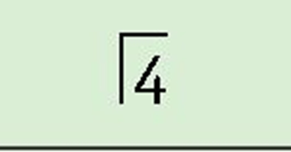
The mesiodistal and faciolingual axial inclinations of the teeth are usually described in terms of angles between the long axis of a tooth and a line drawn perpendicular to a horizontal plane or to the median plane. The axial inclinations of the teeth and roots vary, but in general, they appear to correspond favorably to the data summarized in Box 16-1, adapted from Andrews39 for crowns and consistent with the data of Dempster et al36 for the roots of teeth illustrated in Figure 16-19.
Figure 16-20 A, Anterior and lateral view of average arrangement of the teeth showing mean inclination. Longitudinal axes of roots have been extended beyond the crowns and appear in orthographic projection. B, Orthographic projection of mandibular teeth seen from an anterior and lateral perspective. Root axes are extended beyond the crowns. The degree of obliquity of roots represents an average of the skulls examined.
16-1 Angulation/Inclination of the Teeth*
|
Mesiodistal |
|
|
|
|
|
|
|
|
|
|
Faciolingual |
||
 |
 |
 |
 |
 |
 |
 |
 |
 |
 |
 |
 |
 |
 |
|
8° |
10° |
5° |
9° |
17° |
7° |
2° |
28° |
26° |
16° |
5° |
6° |
8° |
10° |
|
14° |
10° |
9° |
6° |
6° |
0° |
2° |
22° |
23° |
12° |
9° |
9° |
20° |
20° |
 |
 |
 |
 |
 |
 |
 |
 |
 |
 |
 |
 |
 |
 |
*Zsigmondy/Palmer notation system for dentition.
FUNCTIONAL FORM OF THE TEETH AT INCISAL AND OCCLUSAL THIRDS
The incisal and occlusal thirds of the tooth crowns present convex or concave surfaces at all contacting occlusal areas. When the teeth of one jaw come into occlusal contact with their antagonists in the opposite jaw during various mandibular movements, curved surfaces come into contact with curved surfaces. These curved surfaces may be convex or concave. A convex surface representing a segment of the occlusal third of one tooth may come into contact with a convex or a concave segment of another tooth; always, however, curved segments contact curved segments, large or small (Figure 16-21). Lingual surfaces of maxillary incisors present some concave surfaces where convex portions of the incisal ridges of mandibular incisors come into occlu-sal contact.
The posterior teeth show depressions in the depth of sulci and developmental grooves; nevertheless, the enamel sides of the sulci are formed by convexities that point into the developmental grooves.
Figure 16-21 Incisal or occlusal thirds of the crowns of teeth showing curves and concave surfaces of all contacting occlusal areas.
Cusps that are rather pointed contact the rolls of hard enamel that make up marginal ridges on posterior teeth. Until the cusps are worn flat, the deeper portions of the sulci and grooves act as escapements for food, because the convex surfaces of opposing teeth are prevented from fitting into them perfectly by the curved sides of the sulci (Figure 16-22).
Figure 16-22 Maxillary second molar. Note rounded cusp tips and ridges and the rounded, turned "irregular" ridge folding into the central fossa and into the mesial and distal fossae.
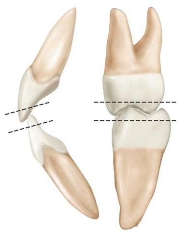
Although the teeth when in centric occlusion seem to intercuspate rather closely, on examination, it is found that escapements have been provided. These escapement spaces are needed for efficient occlusion during mastication. When occluding surfaces come together, some escapement spaces are so slight that light is scarcely admitted through them; they vary in degree of opening from such small ones to generous ones of a millimeter or more at the widest points of embrasure. It should not be assumed that the teeth fit snugly together like gears, at least not unless they are worn down severely from abrasive food or bruxism. As can be seen in Figure 16-23, in unworn teeth the rounded surfaces do not mesh closely together. Escapement space is provided in the teeth by the form of the cusps and ridges, the sulci and developmental grooves, and the interdental spaces or embrasures when teeth come together in occlusion.
The significance of the incisal and occlusal thirds of the teeth has been an area of controversy because so little objective information is available on the influence of flat or convex surfaces on function. The use of anatomical and nonana-tomical occlusal surfaces of denture teeth has only heightened interest in the role of cuspal form, escapements for food, and cutting surfaces in mastication. An examination of contemporary Eskimo or aboriginal dentitions shows that by the age of 20 most of the teeth are flattened, and it has been suggested that the only advantage of cusps on the teeth in humans is the establishment of the teeth in their correct position during the development of occlusion.40 Although such ideas have little acceptance, some clinicians erroneously grind the posterior teeth flat under the impression that dental caries, as well as some forms of TMJ muscle dysfunction and perhaps periodontal disease, may be reduced. Although the evaluation of mastication is an interesting subject and the persistence of cusps into early middle age has occurred only in Western man for the last 200 to 300 years,41,42 it must be accepted that cusp form and function at this time have to be consistent with the dentition of modern man. Although a number of concepts of occlusal morphology have been suggested for restorations, none has been demonstrated to increase the health and comfort of patients more than another.
Figure 16-23 First molars positioned in simulated contact relations in the intercuspal position. In the left illustration the teeth are viewed from a right angle, which suggests that the mesiobuccal cusp of the maxillary molar fits snugly into the mesiobuccal groove of the mandibular molar. However, in the right illustration the view of the tipped molars shows that the triangular ridge of the mesiobuccal cusp does not fit tightly into the mesiobuccal groove and that there are many escapement spaces present.

