The Crown and Root
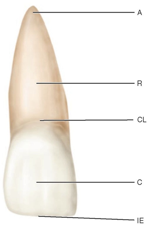
Each tooth has a crown and root portion. The crown is covered with enamel, and the root portion is covered with cementum. The crown and root join at the cementoenamel junction (CEJ). This junction, also called the cervical line (Figure 1-3), is plainly visible on a specimen tooth. The main bulk of the tooth is composed of dentin, which is clear in a cross section of the tooth. This cross section displays a pulp chamber and a pulp canal, which normally contain the pulp tissue. The pulp chamber is in the crown portion mainly, and the pulp canal is in the root (Figure 1-4).The spaces are continuous with each other and are spoken of collectively as the pulp cavity.
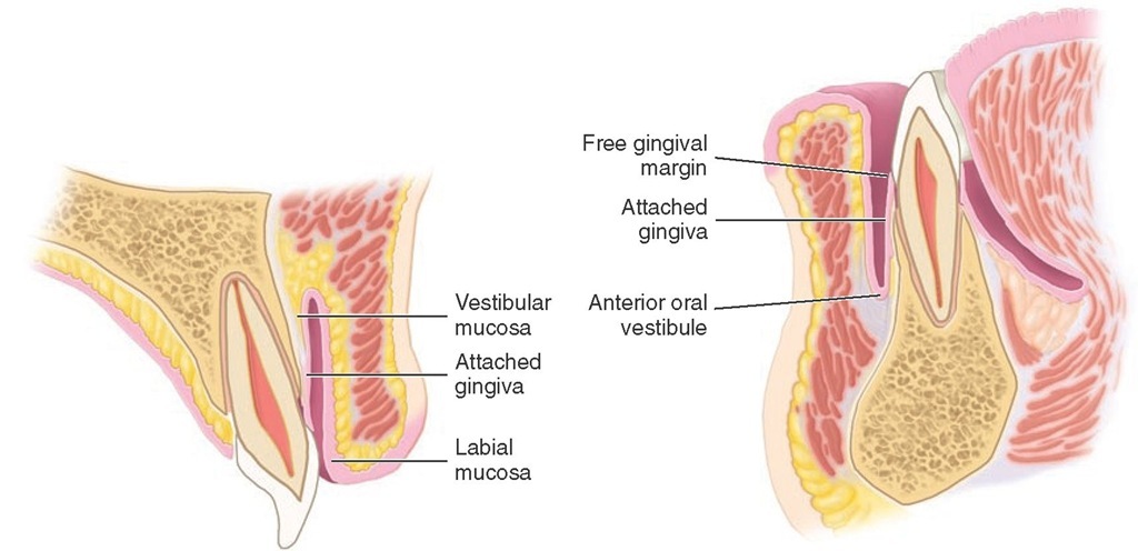
The four tooth tissues are enamel, cementum, dentin, and pulp. The first three are known as hard tissues, the last as soft tissue. The pulp tissue furnishes the blood and nerve supply to the tooth. The tissues of the teeth must be considered in relation to the other tissues of the orofacial structures (Figures 1-5 and 1-6) if the physiology of the teeth is to be understood.
The crown of an incisor tooth may have an incisal ridge or edge, as in the central and lateral incisors; a single cusp, as in the canines; or two or more cusps, as on premolars and molars. Incisal ridges and cusps form the cutting surfaces on tooth crowns.
The root portion of the tooth may be single, with one apex or terminal end, as usually found in anterior teeth and some of the premolars; or multiple, with a bifurcation or trifurcation dividing the root portion into two or more extensions or roots with their apices or terminal ends, as found on all molars and in some premolars.
Figure 1-3 Maxillary central incisor (facial aspect).
The root portion of the tooth is firmly fixed in the bony process of the jaw, so that each tooth is held in its position relative to the others in the dental arch. That portion of the jaw serving as support for the tooth is called the alveolar process. The bone of the tooth socket is called the alveolus (plural, alveoli) (Figure 1-7).
The crown portion is never covered by bone tissue after it is fully erupted, but it is partly covered at the cervical third in young adults by soft tissue of the mouth known as the gingiva or gingival tissue, or "gums." In some persons, all of the enamel and frequently some cervical cementum may not be covered by the gingiva.
Surfaces and Ridges
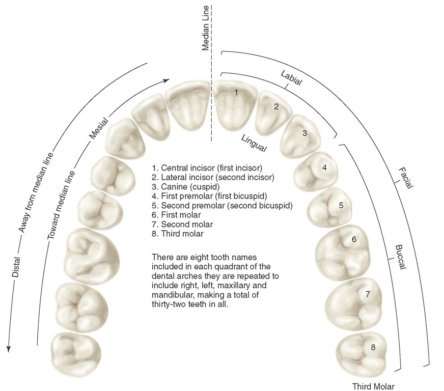
The crowns of the incisors and canines have four surfaces and a ridge, and the crowns of the premolars and molars have five surfaces. The surfaces are named according to their positions and uses (Figure 1-8). In the incisors and canines, the surfaces toward the lips are called labial surfaces; in the premolars and molars, those facing the cheek are the buccal surfaces. When labial and buccal surfaces are spoken of collectively, they are called facial surfaces. All surfaces facing toward the tongue are called lingual surfaces. The surfaces of the premolars and molars that come in contact (occlusion) with those in the opposite jaw during the act of closure are called occlusal surfaces. These are called incisal surfaces with respect to incisors and canines.
Figure 1-4 Schematic drawings of longitudinal sections of an anterior and a posterior tooth. A, Anterior tooth. A, Apex; AF, apical foramen; SC, supplementary canal; B, bone; C, cementum; PM, periodontal ligament; PC, pulp canal; G, gingiva; GC, gingival crevice; GM, gingival margin; PCH, pulp chamber; D, dentin; E, enamel; CR, crown. B, Posterior tooth. A, Apices; PC, pulp canal; PCH, pulp chamber; PH, pulp horn; F, fissure; CU, cusp; CEJ, cementoenamel junction; BI, bifurcation of roots.
Figure 1-5 Sagittal sections through the maxillary and mandibular central incisors.
Figure 1-6 Section through the second maxillary molar and adjacent tissues.
The surfaces of the teeth facing toward adjoining teeth in the same dental arch are called proximal or proximate surfaces. The proximal surfaces may be called either mesial or distal. These terms have special reference to the position of the surface relative to the median line of the face. This line is drawn vertically through the center of the face, passing between the central incisors at their point of contact with each other in both the maxilla and the mandible. Those proximal surfaces that, following the curve of the arch, are faced toward the median line are called mesial surfaces, and those most distant from the median line are called distal surfaces.
Four teeth have mesial surfaces that contact each other: the maxillary and mandibular central incisors. In all other instances, the mesial surface of one tooth contacts the distal surface of its neighbor, except for the distal surfaces of third molars of permanent teeth and distal surfaces of second molars in deciduous teeth, which have no teeth distal to them. The area of the mesial or distal surface of a tooth that touches its neighbor in the arch is called the contact area.
Figure 1-7 Left maxillary bone showing the alveolar process with three molars in place and the alveoli of the central incisor, lateral incisor, canine, and first and second premolars. Note the opening at the bottom of the canine alveolus, an opening that accommodates the nutrient blood and nerve supply to the tooth in life. Although they do not show up in the photograph, the other alveoli present the same arrangement.
Central and lateral incisors and canines as a group are called anterior teeth; premolars and molars as a group, posterior teeth.
Other Landmarks
To study an individual tooth intelligently, one should recognize all landmarks of importance by name. Therefore, at this point it is necessary to become familiar with additional terms, such as the following:
Figure 1-8 Application of nomenclature. Tooth numbers l1 to L8 indicating left maxillary teeth. Tooth surfaces related to the tongue (lingual), cheek (buccal), lips (labial), and face (facial), apply to four quadrants and the upper left quadrant. The teeth or their parts or surfaces may be described as being away from the midline (distal) or toward the midline (mesial).
|
cusp |
triangular ridge |
developmental groove |
|
tubercle |
transverse ridge |
supplemental groove |
|
cingulum |
oblique ridge |
pit |
|
ridge |
fossa |
lobe |
|
marginal ridge |
sulcus |
A cusp is an elevation or mound on the crown portion of a tooth making up a divisional part of the occlusal surface (Figures 1-4 and 1-9).
A tubercle is a smaller elevation on some portion of the crown produced by an extra formation of enamel (see Figure 4-14, A). These are deviations from the typical form.
A cingulum (Latin word for "girdle") is the lingual lobe of an anterior tooth. It makes up the bulk of the cervical third of the lingual surface. Its convexity mesiodistally resembles a girdle encircling the lingual surface at the cervical third (see Figures 1-10 and 4-13, A).
A ridge is any linear elevation on the surface of a tooth and is named according to its location (e.g., buccal ridge, incisal ridge, marginal ridge).
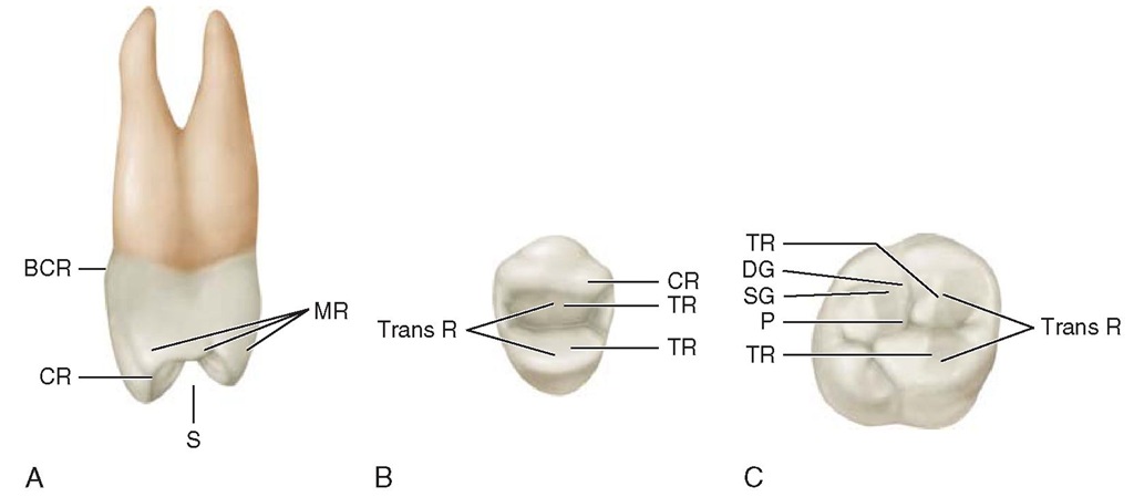
Marginal ridges are those rounded borders of the enamel that form the mesial and distal margins of the occlusal surfaces of premolars and molars and the mesial and distal margins of the lingual surfaces of the incisors and canines (Figures 1-10, A, and 1-11).
FIGURE 1-9 Some landmarks on the maxillary first molar. BCR, Buccocervical ridge; BG, buccal groove; MBC, mesiobuccal cusp; SG, supplemental groove; TF, triangular fossa; MLC, mesiolingual cusp; DG, developmental groove; DLC, distolingual cusp; OR, oblique ridge; DMR, distal marginal ridge; DBC, distobuccal cusp; CF, central fossa.
FIGURE 1-10 A, Maxillary right lateral incisor (lingual aspect). CL, Cervical line;CI, cingulum (also called the linguocervical rìdge); MR, marginal ridge; IR, incisal ridge; LF, lingual fossa. B, Mamelons on erupting, noncontacting central incisors.
FIGURE 1-11 A, Mesial view of a maxillary right first premolar. MR, Marginal ridge;5, sulcus traversing occlusal surface;CR, cusp ridge;ßCR, buccocervical ridge. B, Occlusal view of maxillary right first premolar. CR, Cusp ridge; TR, triangular ridges; Trans R, transverse ridge, formed by two triangular ridges that cross the tooth transversely. C, Occlusal view of a maxillary right first molar. Trans R, Transverse ridge; TR, triangular ridge; P, pit formed by junction of developmental grooves; 5G, supplemental groove; DG, developmental groove; TR, triangular ridge.
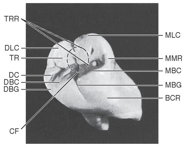
Triangular ridges descend from the tips of the cusps of molars and premolars toward the central part of the occlusal surfaces. They are so named because the slopes of each side of the ridge are inclined to resemble two sides of a triangle (Figures 1-11, B and C, and 1-12). They are named after the cusps to which they belong, for example, the triangular ridge of the buccal cusp of the maxillary first premolar.
When a buccal and a lingual triangular ridge join, they form a transverse ridge. A transverse ridge is the union of two triangular ridges crossing transversely the surface of a posterior tooth (Figure 1-11, B and C).
The oblique ridge is a ridge crossing obliquely the occlusal surfaces of maxillary molars and formed by the union of the triangular ridge of the distobuccal cusp and the distal cusp ridge of the mesiolingual cusp (Figure 1-9).
Central fossae are on the occlusal surface of molars. They are formed by the convergence of ridges terminating at a central point in the bottom of the depression where there is a junction of grooves (Figure 1-12). Triangular fossae are found on molars and premolars on the occlusal surfaces mesial or distal to marginal ridges (Figure 1-9). They are sometimes found on the lingual surfaces of maxillary incisors at the edge of the lingual fossae where the marginal ridges and the cingulum meet (see Figure 4-14, A).
A sulcus is a long depression or valley in the surface of a tooth between ridges and cusps, the inclines of which meet at an angle. A sulcus has a developmental groove at the junction of its inclines. (The term sulcus should not be confused with the term groove.)
A developmental groove is a shallow groove or line between the primary parts of the crown or root. A supplemental groove, less distinct, is also a shallow linear depression on the surface of a tooth, but it is supplemental to a developmental groove and does not mark the junction of primary parts. Buccal and lingual grooves are developmental grooves found on the Pits are small pinpoint depressions located at the junction of developmental grooves or at terminals of those grooves. For instance, central pit is a term used to describe a landmark in the central fossa of molars where developmental grooves join (Figure 1-11, C).
FIGURE 1-12 Mandibular right first molar. MLC, Mesiolingual cusp; MMR, mesial marginal ridge; MBC, mesiobuccal cusp; MBG, mesiobuccal groove; BCR, buccocervical ridge; CF, central fossa; DBG, distobuccal groove; DBC, distobuccal cusp; DC, distal cusp; TR, triangular ridge; DLC, distolingual cusp; TRR, transverse ridge.
A lobe is one of the primary sections of formation in the development of the crown. Cusps and mamelons are representative of lobes. A mamelon is any one of the three rounded protuberances found on the incisal ridges of newly erupted incisor teeth (Figure 1-10, B). (For further description of lobes, see Figures 4-11 through 4-14).
The roots of the teeth may be single or multiple. Both maxillary and mandibular anterior teeth have only one root each. Mandibular first and second premolars and the maxillary second premolar are single rooted, but the maxillary first premolar has two roots in most cases, one buccal and one lingual. Maxillary molars have three roots, one mesiobuc-cal, one distobuccal, and one lingual. Mandibular molars have two roots, one mesial and one distal. It must be understood that description in anatomy can never follow a hard-and-fast rule. Variations frequently occur. This is especially true regarding tooth roots, for example, facial and lingual roots of the mandibular canine.
