Learning Objectives
1. Identify the location of the receptors for each of the five senses and explain how the brain interprets the stimulus for each sense.
2. Describe the major structures of the eye and the functions of each; differentiate between aqueous and vitreous humor.
3. Identify which cranial nerves are responsible for the blink reflex, pupillary changes, visualization, and pain sensation in the eye.
4. Differentiate between myopia, hyperopia, and astigmatism.
5. Identify the structures and dividing lines between the outer, middle, and inner ear.
6. Trace the path of light rays as they enter the eye and focus on the retina; describe transmission to the brain.
7. Trace the path of sound waves through the external, middle, and inner ear. Describe the amplification and interpretation of sound waves.
8. Explain how cerumen, the ossicles, and the eustachian tubes work in protecting the ear.
9. Discuss how the organs of the inner ear work to provide a sense of balance.
10. Identify the location of each type of taste bud on the tongue.
11. Describe the effects of aging on the sensory system.
|
IMPORTANT TERMINOLOGY |
||
|
accommodation |
lacrimal gland |
presbyopia |
|
aqueous humor |
lens |
proprioceptor |
|
astigmatism |
malleus |
ptosis |
|
auricle |
membranous labyrinth |
pupil |
|
cochlea |
myopia |
retina |
|
cones |
olfaction |
rods |
|
conjunctiva |
ophthalmology |
sclera |
|
cornea |
optic disk |
semicircular canal |
|
eustachian tube |
orbit |
stapes |
|
gustation |
organ of Corti |
tinnitus |
|
hyperopia |
ossicle |
tympanic membrane |
|
incus |
otology |
vertigo |
|
iris |
pinna |
vitreous humor |
|
labyrinth |
presbycusis |
|
Without the sensory system, you would know nothing about the world around you. The sensory perceptions are those of seeing, hearing, tasting, smelling, and touching. Humans also receive impressions of warmth, softness, pressure, and pain through the sensory system. Another important perception provided by the sensory system is the sense of equilibrium; that is, knowing whether or not the body is moving and sensing the body’s posture and position. By detecting environmental changes, the sensory system provides humans with protection and with mechanisms for experiencing the world.
Structure and Function
From the study of the nervous system,it is apparent that in order to be aware of information from the world, a person must have the following:
• Receptors to receive a stimulus
• Nerve routes to carry the stimulus to the brain
• Centers in the brain to interpret the stimulus
These principles apply to the sensory system as well. The organs of the sensory system are the eyes, ears, tongue, nose, and skin. The activities of these organs are the senses of vision, hearing, taste, smell, and touch. These senses supply information to the brain and inner body. By providing information, they help the body to detect environmental changes. The body then responds, thereby maintaining homeostasis. Box 21-1 outlines the functions of the sensory system.
The senses of taste, smell, and touch are as interesting and as useful as the senses of sight and hearing. They are not as often involved in illness or disease as are the senses of sight and hearing; however, alterations in these other senses can pose safety hazards. For example, a person whose sense of taste is altered is at risk for ingesting spoiled food. A person whose sense of smell is compromised may not be able to smell smoke. Someone whose sense of touch is altered may not be able to detect hot water and is therefore at risk for burns.The nose is discussed, with the respiratory system.The sense of touch is related to the integumentary system.
Key Concept Sight and hearing are most frequently involved in illness; however; alterations in taste, smell, and touch can present safety hazards. These senses also provide pleasurable sensations.
THE EYE
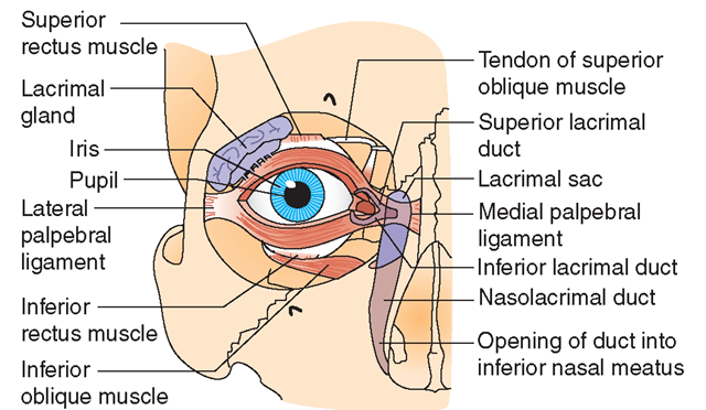
The eye (denoted by the medical prefix ophthalm[o]) is the organ of vision. It lies in a ball-shaped cavity of the skull called the orbit. Figure 21-1 illustrates the major structures of the eye. The medical specialty related to the study of the eye and vision is ophthalmology.
BOX 21-1.
Functions of the Sensory System
♦ Visual sense receives images (light).
♦ Hearing receptors process sound waves (auditory sense).
♦ Through proprioceptors and the inner ear; the system helps to maintain a sense of balance, equilibrium, and position in space.
♦ Chemoreceptors in the mouth obtain information about tastes (gustatory sense).
♦ Chemoreceptors in the nose receive sensations of odors (olfactory sense).
♦ Touch receptors receive information about the surrounding world (e.g., touch, pressure, hot, cold, pain).
♦ Internal organs receive sensations of pain, pressure, fullness.
♦ The brain interprets most of these sensations.
FIGURE 21-1 · The right eye and its appendages (anterior view).
Key Concept The human eye has a 200-degree viewing angle and can see 2.7 million different colors.
The eyelids, or palpebrae, are retractable covers for the eye’s anterior surface. (The medical prefix for eyelid is blephar[o]-). The oval opening between the upper and lower eyelids is called the palpebral fissure. Also covering the anterior eye, beneath and lining the eyelids (the cornea and sclera), is a transparent mucous membrane called the conjunctiva, which is supplied with blood vessels and nerve endings.
The lacrimal glands, which produce tears, keep the eye’s surface moist and lubricated (see Fig. 21-1). Normally, the body produces about 1 mL of lacrimal fluid each day. The lacrimal glands are located at the outer edge (or lateral can-thus) of each eye’s corner. Tears drain out through a small opening, called the nasolacrimal duct, located in the inner corner of the eye (or medial canthus). The nasolacrimal ducts drain these tears into the nose. Tears protect the eyes from infections and foreign objects. Chemical and mechanical irritants (e.g., onions or dust) cause oversecretion by the lacrimal glands to wash the irritants away. (Humans are the only species that form tears in response to emotions.)
Eyeball
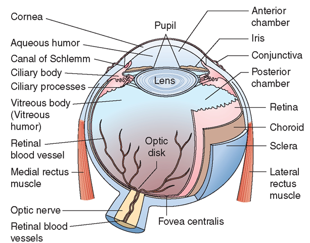
The eyeball is a hollow sphere that consists of three layers of tissue known as tunics: the sclera and cornea, choroid layer, and retina. The lens is another important part of the eyeball, which is illustrated in Figure 21-2.
Sclera and Cornea. The tough protective outer layer of the eyeball is the sclera (the “white” of the eye). The sclera helps to maintain the shape of the eyeball. To permit light rays to enter the front of the eye, the sclera is connected to a transparent, yet tough, section over the front of the eyeball called the cornea. The cornea is covered with stratified squamous epithelium, which is a transparent protective coating. It is one of the structures through which light rays pass. It influences visual acuity by refracting light rays. The cornea is very sensitive to touch and pain. Even minor irritations will stimulate a blinking reflex or a pain sensation.
FIGURE 21-2 · Transverse section of the eyeball.
Protection. The eye is surrounded by the protective bony eye socket which includes a protective cushion of fat. The eyebrows, eyelids, and eyelashes also provide protection. The blink reflex protects the eye from foreign objects or blows. The protective function of tears is described above.
Key Concept The cornea is often removed after death as a tissue for transplantation. Corneas were the first tissues used for transplantation. Transplants restore vision each year to thousands of people with defective or diseased corneas.
Choroid Layer. The middle tunic, the choroid, contains the iris and the ciliary body. This layer, which is vascular, brings oxygen and nutrients to all the layers of the eyes. The colored iris controls the amount of light entering the eye. The ciliary body contains muscles that adjust the shape and thickness of the lens and also secretes aqueous humor (5-6 mL/d), which flows through the anterior chamber of the eye in the space between the cornea and the lens. (The anterior chamber is the space behind the cornea and in front of the iris. The posterior chamber begins behind the iris.) The aqueous humor maintains intraocular (within the eye) pressure (normal is about 24 mm Hg). It also provides nutrients and oxygen to the avascular (without blood vessels) lens and cornea.
Over the front of the eyeball, the choroid develops into a pigmented section, the iris, which gives the eye its specific color. The amount of pigment in the iris determines eye color. The darker the eyes, the more pigment they contain. Lighter eyes, such as blue eyes, contain less pigment. Muscles in the iris control the size of the pupil. The pupil is the black center opening within the eye that allows light to enter the eye. When there is an abundance of light, the muscles of the iris constrict the pupil to allow less light to enter. The reverse occurs under low-light situations (the Purkinje effect).
Special Considerations: LIFESPAN
Eye Color
The iris is normally blue or slate gray in light-skinned infants and brown in dark-skinned infants. Permanent eye color differentiates by age 6 to 9 months.
Lens. The lens is elastic and is located immediately behind the iris. It has a major role in focusing light rays on the retina. The space behind the lens is filled with a transparent gelatin-like material called the vitreous humor. The vitreous humor helps maintain the eyeball’s shape and contributes to intraocular pressure. Loss of the vitreous humor causes blindness.
Retina. The eyeball’s inner layer, the retina, is an incredible light-sensitive membrane. The pigmented layer of the retina is composed of simple cuboidal epithelium and contains the receptors of the optic nerve. The retina also contains specialized neurons, called rods and cones, which permit the perception of light, dark, and color. Each retina contains between 100 and 120 million rods, which are dispersed throughout the retina. Because the pupil dilates in dim light, the light strikes all parts of the retina, thereby activating the rods. Because they are suited to dim light, rods are useful in night vision (scotopic vision). The rods receive sensations of black and white and can register shapes, but not colors.
Each retina also contains approximately 6 to 7 million cones, on which color vision depends. The cones are concentrated in the retina’s center and function in daylight and in bright light (photopic vision). They consist of three individual classes and each receives either red, blue, or green light, which are combined to form colors. Cones also add to visual acuity (visual sharpness), but require a significant amount of light. This explains why you see shades of gray, rather than color, in dim light, because only the rods are receiving stimuli (Table 21-1).
Key Concept The adaptation to darkness (the Purkinje effect) depends on good blood flow to the eye. Blood flow is inhibited by vasoconstrictors, including tobacco and alcohol.
"Color blindness” is a difficulty in distinguishing between colors, particularly red and green. It affects 1 in 30 people, most often men.
TABLE 21-1. Functions and Placements of Rods and Cones
|
|
PLACEMENT |
FUNCTION: COLOR |
FUNCTION: VISION |
|
Rods |
Widespread over retina |
Receive black and white and shapes |
Scotopic vision |
|
Cones |
Center of retina |
Receive color |
Photopic vision |
Nerves and Muscles
The optic nerve (cranial nerve II) carries the stimuli for vision from each eye. The stimuli are transmitted from the optic nerve of one eye and meet the optic nerve of the other eye at the optic chiasm. At the optic chiasm, the optic nerves cross, then continue as the optic tract, and then are conducted to the brain’s occipital lobe in the cerebral cortex. The left side of the occipital lobe receives visual images from the right side of an object, whereas the right side of the occipital lobe receives visual images from the left side of an object. The brain’s occipital lobe must translate these images. The ophthalmic nerve, a branch of the trigeminal nerve (cranial nerve V), carries sensations of eye pain and temperature to the brain. For example, if a foreign object is in the eye, the ophthalmic nerve carries that sensation.
Smooth muscles control pupil size and lens action. Three pairs of extraocular (outside the eye) muscles, attached to the sclera, move the eyeball. Another muscle, attached to the upper eyelid, holds the eye open; when this muscle relaxes, the eyelid shuts. The oculomotor nerve (cranial nerve III) innervates some of the voluntary muscles that move the eyeball and eyelid. This cranial nerve is also involved in some autonomic eye reactions, such as pupil accommodation to varying degrees of light. The trochlear nerve (cranial nerve IV) assists with some voluntary eyeball movements. The abducens nerve (cranial nerve VI) coordinates with cranial nerves III and IV to move the eyes (Table 21-2).
Key Concept Both sides of the brain’s occipital lobe interpret visual images. Smooth (involuntary) muscles control pupil size and lens accommodation.
TABLE 21-2. Nerves and Muscles of the Eye
|
CRANIAL NERVE |
FUNCTION |
|
II Optic |
Carries visual images to the brain |
|
III Oculomotor |
Constricts and dilates the pupil; elevates the eyelid; innervates the superior, inferior, and medial rectus, and inferior oblique muscles |
|
IV Trochlear |
Voluntary eye movement |
|
V Trigeminal (ophthalmic branch) |
Carries sensations of eye pain and temperature |
|
VI Abducens |
Innervates the lateral rectus muscle |
|
VII Facial |
Controls the blinking reflex |
|
MUSCLE |
FUNCTION |
|
Superior rectus |
Controls upward movement |
|
Inferior rectus |
Controls downward movement |
|
Lateral rectus |
Controls outward movement |
|
Medial rectus |
Controls inward movement |
|
Superior oblique |
Controls upward and outward movement |
|
Inferior oblique |
Controls downward and inward movement |
THE EAR
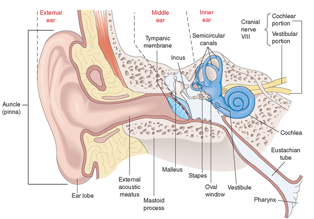
The ear is the organ of hearing and equilibrium. It has three parts: external, middle, and inner ear (Fig. 21-3). The specialty concerned with the ear and hearing is otology.
FIGURE 21-3 · The ear; with its external, middle, and inner subdivisions.
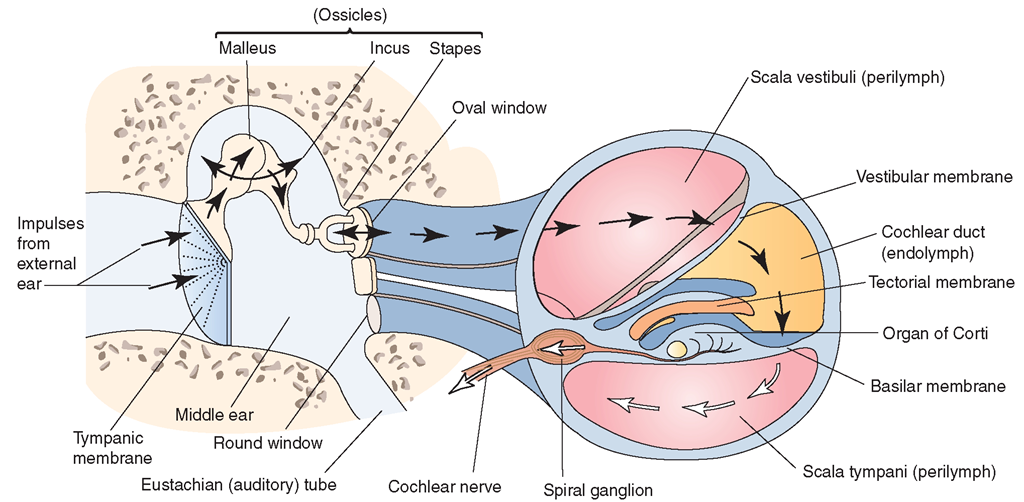
FIGURE 21-4 · Path taken by sound waves reaching the inner ear Sound travels from the external environment s external auditory canal s tympanic membrane, where vibrations begin. The vibrations travel through the middle ear s oval window into the inner ear, where they travel through the cochlear fluid (perilymph and endolymph) s receptors (hair cells) of organ of Corti. From here, the vibrations are transmitted to auditory nerve fibers s vestibulocochlear nerve s cerebral cortex, where they are interpreted.


