Herniation of the Brain
When ICP exerts enough pressure to displace a portion of the brain, herniation (an upward, downward, or lateral pushing of a portion of the brain through an opening) can occur. This opening can be a natural intracranial opening, such as the foramen magnum. The brain herniates (pushes) through the large foramen (opening) in the occipital bone, which lies between the cranial and spinal cavities.
FIGURE 78-6 · Monitoring intracranial pressure. (A) Fiberoptic, transducer-tipped pressure and temperature ventricular monitoring catheter. (B) Ventricular bolt that is connected to a pressure transducer and display system.
Herniation can also occur through a previous craniotomy site or through an opening caused by trauma. Herniation causes severe injury to the brain because of prolonged hypoxia to parts of the brain that control the vital functions of the body—breathing and blood circulation. The result is brain death and death of the individual.
When ICP is elevated, an LP is contraindicated because the withdrawal of even a small amount of CSF can cause the brain to shift, or herniate. Therefore, a safer method of determining ICP is ICP monitoring.
Concussion
A concussion is the result of any blow to the head. The concussion may not damage any brain structures, but temporary unconsciousness is possible. The length of time a person remains unconscious varies. Some clients recover from concussions with no apparent ill effects except the inability to remember the event; others have blurred vision or severe headaches. A client who has had anything other than a very minor concussion should see a physician immediately for a thorough neurologic examination.
In coup-contrecoup injuries, damage occurs both at and opposite to the site of impact. The brain may be hit on one side (coup) and then bounce (rebound) off the other side of the skull (contrecoup). The brain is partially anchored at the brain stem and floats in CSF within the cranium. With direct and rebound trauma, blood vessels, nerve tracts, brain tissue, and other structures are bruised and torn. Serious injuries may also occur to the brain stem because of the contrecoup action.
After the original injury, a postconcussion syndrome may persist for several weeks to months. Symptoms include headache, anxiety, fatigue, or vertigo (a sensation of rotation of self or one’s surroundings; not true dizziness).
Laceration and Contusion
A laceration is tearing of the brain tissue caused by direct impact or penetrating injury. Lacerations are commonly associated with depressed skull fractures, which are discussed below. In contusion, the brain tissue is bruised.
Skull Fractures
A skull fracture may be open, closed, simple, depressed, or comminuted (fragmented), depending on whether the skull and scalp are intact. Many skull fractures are minor, being no more than cracks in the bone. Usually, they heal without difficulty. Any scalp lacerations must be thoroughly examined to determine if the cranium has been opened.
Open skull fractures potentially expose the brain to external microorganisms, which could lead to meningitis or encephalitis. However, open skull fractures are less likely to produce rapid elevations in ICP. The fracture allows for some brain swelling.
A depressed skull fracture is caused by a severe blow to the head. The fracture breaks the bone and forces the broken edges to press against the brain, resulting in a significant risk for ClCP and meningitis. Effects vary with the injury’s severity and location. If, for example, the bone fragment presses on the brain’s speech center, the client’s speech may be impaired until the pressure is relieved.
A basilar skull fracture is a fracture at the base of the skull. It may injure the nerves entering the spinal cord or interfere with CSF circulation. Basilar skull fractures can tear the dura.
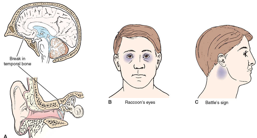
In a basilar skull fracture, rhinorrhea, leakage of CSF from the nose (otorrhea), or leakage of CSF from the ear may occur. The nurse may be asked to test this fluid for the presence of CSF. A positive test for CSF is known as a halo sign (see In Practice: Nursing Care Guidelines 78-3).
Figure 78-7 illustrates the effects of a basilar fracture with periorbital ecchymosis (raccoon’s eyes) and periauricular ecchymosis (Battle’s sign). A basilar skull fracture is especially dangerous because of potential damage to the vital centers that control blood pressure and respiration.
Hematoma
A hematoma refers to a blood clot within the skull. Hypertension and trauma are the most common causes. With any type of cranial hematoma, ICP may dangerously elevate.
IN PRACTICE :NURSING CARE GUIDELINES 78-3
DETERMINING CEREBROSPINAL FLUID (CSF)IN DRAINAGE
• Wet a chemical reagent strip, such as a Dextrostix, with drainage from the nose or ear.
• Observe the color change and whether it indicates the presence of glucose. The presence of glucose in the fluid suggests that the fluid is CSF; test is positive.
If the test is positive and there also is blood (which also contains glucose) in the drainage:
• Collect droplets of drainage on a white absorbent pad.
• Inspect the wet area after a few minutes for a halo sign: If a yellow ring encircles a central ring that is red, the red ring indicates blood, and the yellow ring suggests CSF.
Halo sign. Clear drainage that separates from bloody drainage suggests the presence of CSF.
The swelling or mass of blood compresses brain tissue, creating further damage. Herniation of brain tissue is possible. Carefully observe the client for any signs of tICP. Specific signs and symptoms are determined by the area of the brain affected and the extent of any neurologic damage.
Epidural Hematoma
An epidural hematoma is an accumulation of blood, usually from the temporal artery, between the dura and the skull (Fig. 78-8A). The pressure of an epidural hematoma can quickly cause seizures, paralysis, and death. One or both of the person’s pupils may be dilated. Usually, the person is unconscious immediately after the injury, lucid for a brief period, then unconscious again as blood accumulates in the epidural space and causes pressure. Epidural hematomas are most common in children. The mechanism of injury is typically a blow to the side of the head.
FIGURE 78-7 · (A) Basilar skull fracture in the temporal bone can cause cerebrospinal fluid (CSF) to leak from the nose or ear (B) Periorbital ecchymosis, called raccoon’s eyes. (C) Battle’s sign over the mastoid process.
FIGURE 78-8 · (A) Epidural hematoma. (B) Intracranial hematoma. (C) Subdural hematoma.
Intracranial Hematoma
An intracranial (intracerebral) hematoma is caused by hemorrhage and edema that results from bleeding within the skull (Fig. 78-8B). The cause may be rupture of delicate blood vessels owing to hypertension or a cerebral aneurysm. Ruptured blood vessels within the brain are one cause of CVAs.


