The Photoreceptors
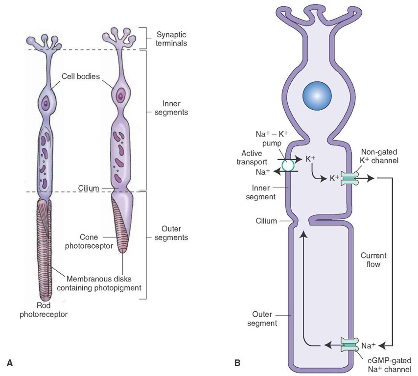
As noted earlier, the human retina consists of two types of photoreceptors: the rods and cones. The rods and cones consist of the following functional regions: an outer segment, an inner segment, and a synaptic terminal (Fig. 16-2A).
The outer segment is located toward the outer surface of the retina and is involved in phototransduction (described later). This segment consists of a stack of membranous discs that contain light-absorbing photo-pigments. These discs are formed by an infolding of the plasma membrane. In the rods, these discs are free floating because they pinch off from the plasma membrane. In the cones, the discs remain attached to the plasma membrane. The outer segments are constantly being renewed. The discarded tips are removed by phagocytosis by pigment epithelial cells. The inner segment contains the nucleus and most of the biosynthetic mechanisms. The inner segment is connected to the outer segment by a stalk or cilium that contains microtubules. The synaptic terminal makes synaptic contact with the other cells.
Cones
Cones are responsible for daylight vision. Vision mediated by cones is of higher acuity than that mediated by rods. Cones mediate color vision, whereas rods do not. Cones have a fast response, and their integration time is short. Their conical shape makes them more sensitive to direct axial rays. As mentioned earlier, they are concentrated in the fovea. The loss of cones results in decreased visual acuity and color vision deficits. Individuals with loss of cones are considered to be legally blind.
Rods
Rods are highly sensitive and can detect dim light. They are specialized for night vision and saturate in daylight. The loss of rods results in night blindness and loss of peripheral vision. They contain more photosensitive pigment than the cones. The photosensitive pigment is responsible for the ability of rods to capture more light.
Both rods and cones, unlike ganglion cells, do not respond to light with an action potential. Instead, they respond with graded changes in membrane potential. The response of rods is slow, whereas the response of cones is fast (Table 16-1).
Phototransduction
In the outer segment membrane of the photoreceptors (rods and cones), there are cyclic guanosine monophosphate (cGMP)-gated Na+ (sodium) channels. cGMP binds directly to the cytoplasmic side of the channel, which causes it to open, allowing an influx of Na+. During darkness, the presence of high levels of cGMP in photoreceptors results in opening of Na+ channels, and an inward current carried by Na+ flows into the outer segment of the photoreceptor (Fig. 16-2B). Thus, the photoreceptors remain depolarized during darkness. K+ (potassium) flows out across the inner segment of the receptor membrane through nongated K+ (leakage) channels. Steady intracellular concentrations of Na+ and K+ are maintained by Na+-K+ pumps located in the inner segments of the photoreceptor.
FIGURE 16-2 The structure of the photoreceptors. (A) Each type of photoreceptor (rods and cones) consists of three regions: an outer segment, an inner segment, and the region of synaptic terminals. (B) Note the location of cGMP-gated Na+ channels and nongated K+ (leakage) channels. For details, see text. cGMP = cyclic guanosine monophosphate; Na+ = sodium; K+ = potassium.
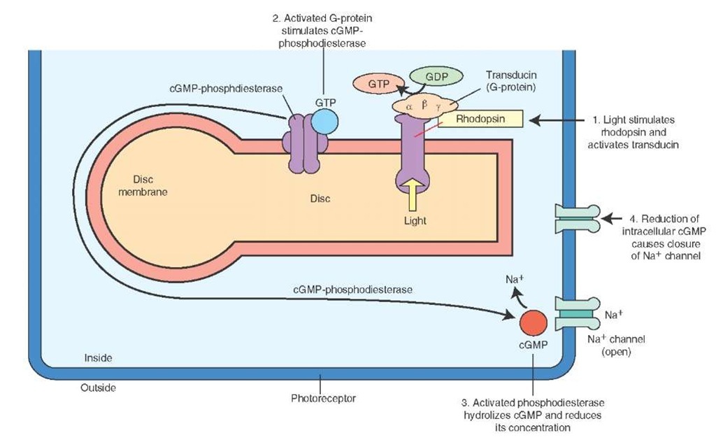
FIGURE 16-3 The mechanism of phototransduction in rods. The retinal component of rhodopsin (photopigment of rods) absorbs light, the conformation of rhodopsin is changed, a G protein (transducin in rods) is activated, cGMP phosphodiesterase is activated, hydrolysis of cGMP takes place, reducing its concentration, cGMP-gated Na+ channels are closed, the influx of intracellular Na+ is reduced, and the photoreceptor cell is hyperpolarized. cGMP = cyclic guanosine monophosphate; Na+ = sodium; GTP = guanosine triphosphate; GDP = guanosine diphosphate.
A photoreceptor pigment, rhodopsin, is present in the rods. It consists of a protein called opsin that is attached with the light-absorbing component, retinal (an aldehyde form of vitamin A). Opsin is embedded in the disc membrane and does not absorb light. In the cones, the protein is called cone-opsin, and it is attached with a light-absorbing component similar to that present in rhodopsin. The events that occur in phototransduction are shown in Figure 16-3: (1) the retinal component of rhodopsin absorbs light, which results in a change in the conformation of the photoreceptor pigment, and a G protein (called transducin in rods) is stimulated; (2) the G protein activates cGMP phosphodiesterase (PDE); (3) the activated PDE hydrolyzes cGMP and reduces its concentration; (4) a reduction in the concentration of cGMP results in closing of the cGMP-gated Na+ channels; and (5) the influx of Na+ is reduced, and the photoreceptor cell is hyperpolarized. Thus, photoreceptors produce a hyperpo-larizing generator (receptor) potential instead of a depolarizing generator potential, which is observed in other receptors. The photoreceptors (rods and cones) do not fire action potentials.
The rods and cones make synaptic contacts with the dendrites of bipolar and horizontal cells. The signals from rods and cones are transmitted to the bipolar and horizontal cells via chemical synapses. As mentioned earlier, vision during normal daylight depends on cones, while night vision involves rods.
Processing of Signals From the Photoreceptors by Different Retinal Cells
Bipolar, Horizontal, and Ganglion Cells
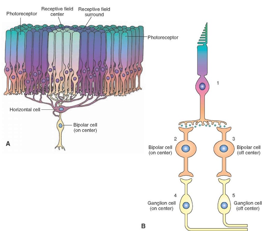
The cell bodies of bipolar neurons are located in the inner nuclear layer of the retina. These cells constitute the main link in the transmission of visual signals from rods and cones to ganglion cells. The receptive field of a bipolar cell is a circular area of the retina that, when stimulated by a light stimulus, changes the membrane potential of the bipolar cell. The receptive field of a bipolar cell consists of two parts: the receptive field center, which provides a direct input from the photoreceptors to the bipolar cells, and the receptive field surround, which provides an indirect input from the photoreceptors to the bipolar cells via horizontal cells (Fig. 16-4A). The changes in membrane potential of bipolar cells to a light stimulus upon the receptive field center and surround are opposite.
The mechanism of membrane potential changes in the bipolar cells in response to light can be summarized as follows. There are two populations of bipolar cells: "on"-center bipolar cells and "off"-center bipolar cells. When stimulated, bipolar cells exhibit graded potentials rather than action potentials.Each photoreceptor cell (e.g., a cone; Fig. 16-4B, 1) synapses on an on-center (Fig. 16-4B, 2) and an off-center bipolar cell (Fig. 16-4B, 3). Each on-center bipolar cell, in turn, synapses with an on-center ganglion cell (Fig. 16-4B, 4), and each off-center bipolar cell synapses with an off-center ganglion cell (Fig. 16-4B, 5).
FIGURE 16-4 Receptive fields of photoreceptors and their connections. (A) The receptive field center provides a direct input from the photoreceptors to the bipolar cell, and the receptive field surround provides indirect input from the photoreceptor to the bipolar cells via horizontal cells. (B) 1: Photoreceptor cell; 2: on-center bipolar cell; 3: off-center bipolar cell; 4: on-center ganglion cell; 5: off-center ganglion cell.
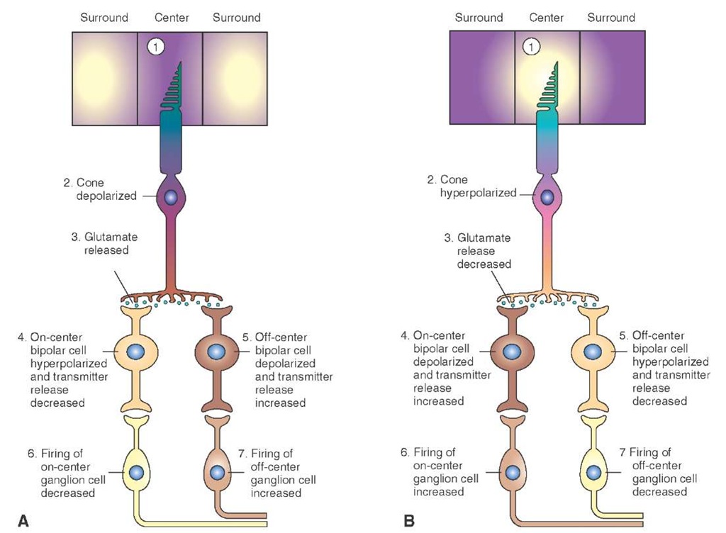
FIGURE 16-5 Responses of retinal bipolar and ganglion cells to darkness and illumination in the receptive field center. (A) Changes in the electrical activity of the photoreceptor and on-center and off-center bipolar and ganglion cells when the photoreceptor receptive field center is in the dark. (B) Changes in the electrical activity of the photoreceptor and on-center and off-center bipolar and ganglion cells when the photoreceptor receptive field center is illuminated.
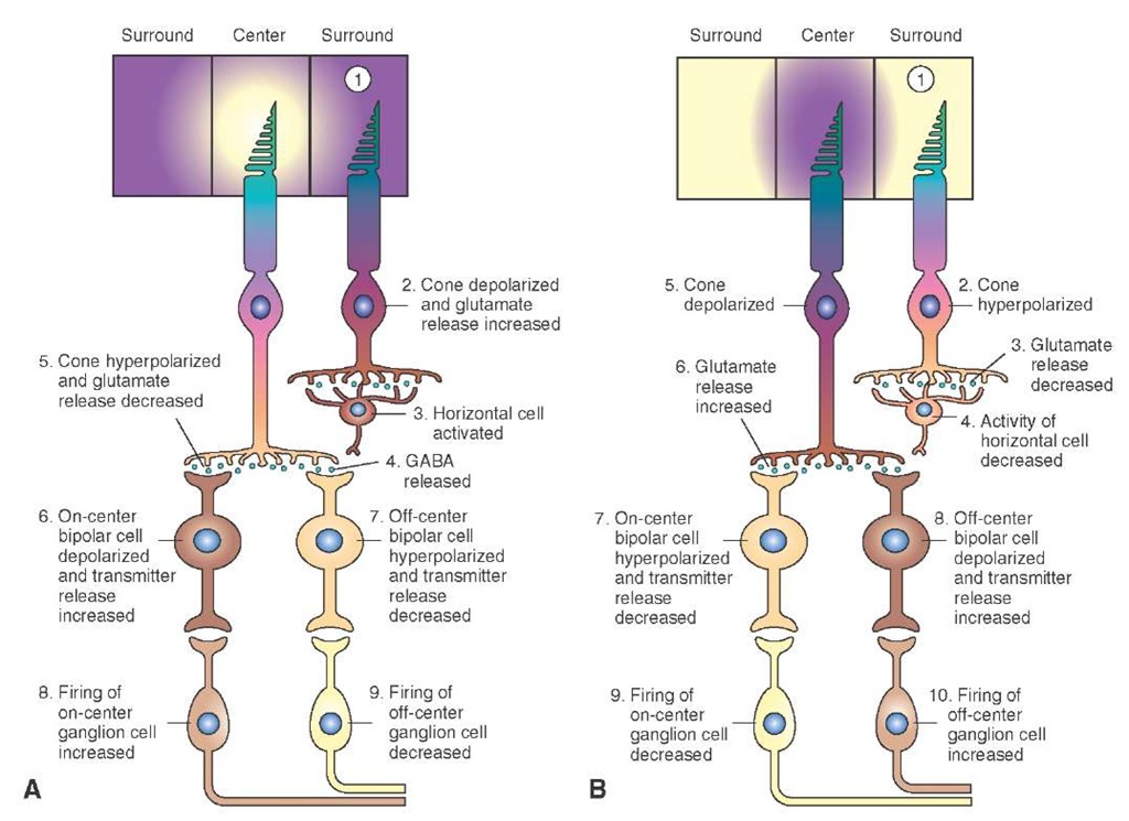
FIGURE 16-6 Responses of retinal bipolar and ganglion cells to darkness and illumination in the receptive field surround. (A) Changes in the electrical activity of the photoreceptor and on-center and off-center bipolar and ganglion cells when the photoreceptor receptive field surround is in the dark. (B) Changes in the electrical activity of the photoreceptor and on-center and off-center bipolar and ganglion cells when the photoreceptor receptive field surround is illuminated. See text for details. GABA = gamma aminobutyric acid.
When the receptive field center is in dark (Fig. 16-5A, 1), the photoreceptors are depolarized (Fig. 16-5A, 2), and they release glutamate constantly (Fig. 16-5A, 3). Glutamate released from the photoreceptor terminals stimulates metabotropic glutamate receptors on the on-center bipolar cells, K+ (potassium) channels are opened, there is an efflux of K+, the on-center bipolar cell is hyperpolarized, and the release of its transmitter (probably glutamate) is decreased (Fig. 16-5A, 4). On the other hand, glutamate released from the photoreceptor terminals stimulates ionotropic glutamate receptors on the off-center bipolar cells, Na+ channels are opened, Na+ flows into the cell, the off-center bipolar cell is depolarized, and the release of its transmitter (probably glutamate) is increased (Fig. 16-5A, 5).
Hyperpolarization of on-center bipolar cells (Fig. 16-5A, 4) results in a decrease in the release of their transmitter, which, in turn, results in a decrease in the firing of the corresponding on-center ganglion cells (Fig. 16-5A, 6). Depolarization of off-center bipolar cells (Fig. 16-5A-5) results in an increase in the release of their transmitter which, in turn, results in an increase in the firing of the corresponding off-center ganglion cells (Fig. 16-5A, 7).
When the photoreceptor in the receptive field center receives a light stimulus (Fig. 16-5B, 1), it is hyperpolarized (Fig. 16-5B, 2), and glutamate release from its terminals is decreased (Fig. 16-5B, 3). The reduction in the release of glutamate from the photoreceptor terminals causes depolarization of the on-center bipolar cell and an increase in its transmitter release (Fig. 16-5B, 4), whereas the off-center bipolar cell is hyperpolarized, and there is a decrease in its transmitter release (Fig. 16-5B, 5). Depolarization of on-center bipolar cells (Fig. 16-5B, 4) results in an increase in the release of their transmitter, which, in turn, results in an increase in the firing of the corresponding on-center ganglion cells (Fig. 16-5B, 6). Hyperpolarization of off-center bipolar cells (Fig. 16-5B, 5) results in a decrease in the release of their transmitter, which, in turn, results in a decrease in the firing of the corresponding off-center ganglion cells (Fig. 16-5B, 7).
Bipolar and ganglion cells elicit opposite responses when light is received at the receptive field surround. The photoreceptors located in the receptive field surround of the bipolar cells are connected to photoreceptors in the receptive field center by interneurons called horizontal cells. During darkness (Fig. 16-6A, 1), the photoreceptors in the receptive field surround are depolarized, and there is an increase in the release of their transmitter (glutamate) (Fig. 16-6A, 2). Glutamate released from the photoreceptors activates horizontal cells (Fig. 16-6A, 3) that, in turn, release an inhibitory transmitter (probably gamma aminobutyric acid [GABA]) (Fig. 16-6A, 4) at their synapses with photoreceptors located in the receptive field center and cause their hyperpolarization and decrease in the glutamate release from their terminals (Fig. 16-6A, 5). This phenomenon is called lateral inhibition. Decrease in glutamate release from the terminals of photoreceptors in the receptive field center results in depolarization of on-center bipolar cells and an increase in the release of their transmitter (Fig. 16-6A, 6), whereas the off-center bipolar cells are hyperpolarized, and there is a decrease in the release of their transmitter (Fig. 16-6A, 7). As mentioned earlier, depolarization of on-center bipolar cells (Fig. 16-6A, 6) causes an increase in the firing of corresponding ganglion cells (Fig. 16-6A, 8), while hyperpolarization of the off-center bipolar cells (Fig. 16-6A, 7) causes a decrease in the firing of corresponding ganglion cells (Fig. 16-6A, 9).
When light is received at the receptive field surround (Fig. 16-6B, 1), the photoreceptor in this field is hyperpolarized (Fig. 16-6B, 2). There is a decrease in the release of glutamate from their terminals (Fig. 16-6B, 3), activity of horizontal cells is decreased (Fig. 16-6B, 4), GABA release at the terminals of horizontal cells is decreased, and lateral inhibition exerted by them on the photoreceptors in the receptive field center is decreased. Thus, the photorecep-tors located in the receptive field center are released from lateral inhibition from horizontal cells. Consequently, the photoreceptors in the receptive field center are depolarized (Fig. 16-6B, 5), and there is an increase in the glutamate release from their terminals (Fig. 16-6B, 6). Subsequently, the on-center bipolar cells are hyperpolarized, and there is a decrease in the release of their transmitter (Fig. 16-6B, 7). The off-center bipolar cells are depolarized, and there is an increase in the release of their transmitter (Fig. 16-6B, 8). Again, hyperpolarization of on-center bipolar cells (Fig. 16-6B, 7) causes a decrease in the firing of corresponding ganglion cells (Fig. 16-6B, 9), whereas depolarization of off-center bipolar cells (Fig. 16-6B, 8) causes an increase in the firing of corresponding ganglion cells (Fig. 16-6B, 10).
