Globus Pallidus
The globus pallidus lies immediately lateral to the internal capsule and medial to the putamen (Fig. 13-12). The globus pallidus consists of two parts: a lateral and a medial segment, which are separated by a thin band of white matter sometimes referred to as medial medullary lamina. A lateral medullary lamina separates the globus pallidus from the putamen. The rostral limit of the globus pallidus extends to a level just beyond the anterior commissure, and the caudal limit is approximately the mid-level of the thalamus. In contrast to the neostriatum, which constitutes the primary receiving area for inputs into the basal ganglia, the globus pallidus represents the primary region for the outflow of information from the basal ganglia. Specifically, fibers, which form the ansa lenticularis and lenticular fasciculus, arise from the globus pallidus (see the following section).
Fiber Pathways of the Basal Ganglia
Two pathways emerge from different parts of the globus pallidus and can be detected in a cross-sectional illustration taken through the middle of the diencephalon (Fig. 13-12). The ansa lenticularis emerges from the medial segment of the globus pallidus and passes in a ventromedial direction and caudally toward the midbrain (Figs. 13-8 and 13-12). The second pathway is called the lenticular fasciculus (Fig. 13-12). This pathway passes in a dorsomedial direction from the pallidum around the dorsal surface of the subthalamic nucleus. The fibers then curve around the dorsal aspect of the zona incerta, passing in a dorsolateral direction caudally toward the midbrain. When both the ansa lenticularis and lenticular fasciculus reach a position immediately rostral to the red nucleus, they reverse their course, turning now in a rostral direction while appearing to merge. These fiber bundles, which are joined by other fibers from the cerebellum called denta-to thalamic fibers, are collectively referred to as the thalamic fasciculus. Fibers from the thalamic fasciculus ultimately terminate mainly in the ventrolateral and ventral anterior thalamic nuclei. The region immediately in front of the red nucleus where fibers of the lenticular fasciculus and ansa lenticularis merge and course rostrally is called the H field of Forel (see Fig. 20-7). The H field of Forel is sometimes referred to as the pre-rubral field, a region that lies immediately rostral to the red nucleus at the dien-cephalic-mesencephalic juncture. The thalamic fasciculus is called the H1 field of Forel, and the lenticular fasciculus is called the H2 field of Forel (Fig. 13-12).
Limbic System and Associated Structures of The Basal Forebrain
The term "limbic system" ("limbic" meaning border) has been used to describe a ring of structures that were originally thought to be associated mainly with olfactory functions and that lie between the neocortex and diencephalon. The primary structures that constitute the limbic system (or limbic structures) include the hippocampal formation, septal area, amygdala, and adjoining regions of cortex (i.e., entorhinal and pyriform cortices). In addition, the prefrontal cortex and cingulate gyrus are often included because of their anatomical and functional relationships with processes normally attributed to the limbic system.
Hippocampal Formation
The hippocampal formation lies deep within the temporal lobe. It is an elongated structure that is oriented in an anterior-to-posterior plane along the longitudinal axis of the temporal lobe. It also forms the medial wall of the inferior horn of the lateral ventricle along its entire length (Fig. 13-13). In its histological appearance, the hippocampus appears as a primitive form of cortex and is, therefore, composed of distinct cell and fiber layers, which bear some resemblance to the neocortex.
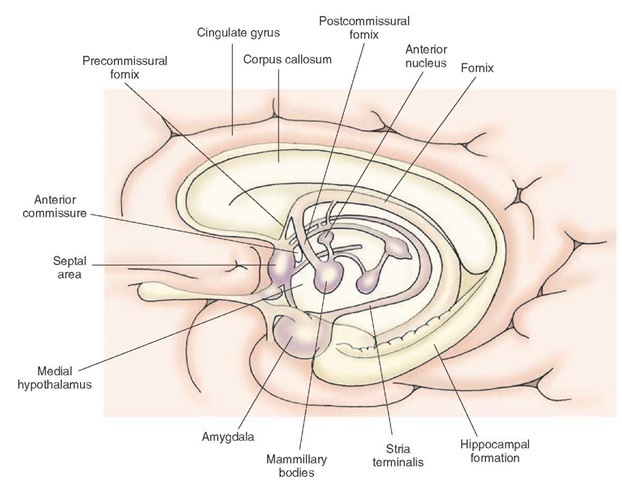
The major outflow pathway of the hippocampal formation is the fornix (Figs. 13-2 and 13-13). The fibers of the fornix initially pass in a dorsomedial direction toward the midline of the brain until they occupy a position immediately below the corpus callosum. The fornix fibers then pass rostrally toward the front of the brain until they approach the level of the anterior commissure. Many of the fornix fibers then pass ventrally behind the anterior commissure into the diencephalon where they terminate in the anterior thalamic nucleus and mammillary bodies of the hypothalamus. The component of the fornix that is distributed to the diencephalon is called the postcommis-sural fornix. Another component of the fornix passes rostral to the anterior commissure and is distributed to the septal area. This component of the fornix is called the precommissural fornix (Fig. 13-13). The hippocampal formation and septal area contribute to the central regulation of emotional behavior, motivational processes, hormonal and autonomic regulation, and memory functions.
Septal Area
The septal area is an important region of the limbic system because it serves, in part, as the relay nucleus of the hippocampal formation to the medial and lateral hypoth-alamus (Fig. 13-14). The septal area is a primary receiving area for fibers contained within the precommissural fornix that arise from the hippocampal formation. The septal area, in turn, projects many of its axons to different regions of the hypothalamus. Accordingly, the septal area may be viewed as a functional extension of the hippocampal formation.
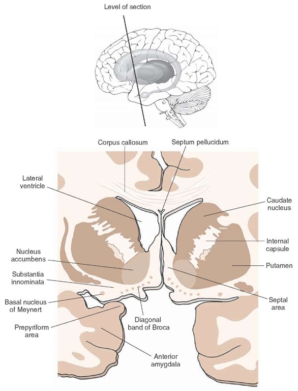
In animals, including nonhuman primates, the septal area consists of two regions: a dorsal and a ventral division (Fig. 13-15). The dorsal division occupies a position in the anterior aspect along the midline of the forebrain rostral to the anterior commissure, thus forming the medial wall of the anterior horn of the lateral ventricle.
FIGURE 13-13 The positions occupied by different components of the limbic system. The amygdala is situated in a rostral position in the temporal lobe, and the hippocampal formation extends caudally from its rostral pole proximal to the amygdala. A major component of the fornix projects to the diencephalon (postcommissural fornix), and another component reaches the septal area (precommissural fornix). The stria terminalis projects from the amygdala to the ventromedial hypothalamus. Olfactory inputs form two fiber bundles: a lateral olfactory stria, which innervates temporal lobe structures, and a medial olfactory stria, which supplies the septal area and contralateral olfactory bulb.
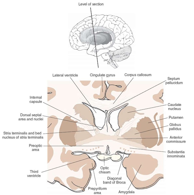
FIGURE 13-14 Cross section taken through the forebrain at the level of the anterior commissure. Note the positions occupied by the septal nuclei, bed nucleus of the stria terminalis, and preoptic region. The inset indicates the plane of the section.
The dorsal septal area in lower forms of animals is cell rich and, therefore, provides a thickness to this structure not seen in higher forms. The ventral region contains many cells of a relatively large size that are situated adjacent to the medial edge of the basal forebrain. Moreover, the cells lie parallel to the medial edge of the brain and extend ventrally to the base of the brain. At the base of the brain, this group of cells then shows a horizontal (or lateral) orientation for a short distance. The cells present within the ventral division of the septal area are referred to as the diagonal band of Broca. In humans, the dorsal septal area is virtually absent and contains only a septum pellucidum, which is devoid of nerve cells. Instead, the septal area is relegated to the ventral region (Fig. 13-15).
There are other cells groups of the basal forebrain that are of functional importance and that lie close to the septal area. These include the bed nucleus of the stria terminalis and several regions of the basal forebrain (the substantia innominata and nucleus accumbens).
Bed Nucleus of the Stria Terminalis
The bed nucleus of the stria terminalis lies in a position immediately ventrolateral to the column of the fornix as it is about to enter the diencephalon at the level of the anterior commissure (Fig. 13-14). It receives many fibers from the amygdala and contributes fibers to the hypotha-lamus and autonomic centers of the brainstem. The stria terminalis is believed to regulate autonomic, endocrine, and affective processes normally associated with the amygdala.
Nucleus Accumbens
The nucleus accumbens is found at the level of the head of the caudate nucleus. Its ventral border is the substantia innominata, the medial border is the septal area, and its lateral and dorsal borders are the putamen and caudate nucleus, respectively (Fig. 13-15). The nucleus accumbens receives a large dopaminergic projection from the brain-stem and other inputs from the amygdala and parts of the hippocampal formation. In turn, it projects its axons to the substantia innominata, substantia nigra, and ventral teg-mental area. The nucleus accumbens is believed to integrate the sequencing of motor responses associated with affective processes. These include functions related to addictive processes as well.
FIGURE 13-15 Cross section taken through the forebrain at a level rostral to that shown in Figure 13-14. Key nuclei of the basal forebrain include the diagonal band of Broca and substantia innominata. The inset indicates the plane of the section.
Substantia Innominata
The substantia innominata, which is sometimes identified as part of the extended amygdala, is located at the level of the septal nuclei immediately below the striatum. Its medial border is the diagonal band of Broca and preoptic region. The substantia innominata extends laterally to a position where its cells appear to merge with those of the prepyriform area. Its ventral border is the base of the brain (Figs. 13-14 and 13-15).
Near the base of the brain can be found a large-celled region called the basal nucleus of Meynert. The unique feature of these cells is that they project their axons to wide regions of the cerebral cortex and to limbic structures, such as the hippocampal formation and amygdala. Moreover, these cells have been shown to be cholinergic. In Alzheimer’s patients, there is a loss of cholinergic inputs to the cerebral cortex coupled with degeneration of these cells, suggesting that damage to the basal nucleus of Meynert may contribute to the etiology of Alzheimer’s disease. The substantia innominata shares reciprocal connections with the amygdala, and projects its axons to the lateral hypothalamus as well. In this manner, the substantia innominata might also serve as a relay of signals from parts of the amygdala to the lateral hypothalamus.
Amygdala
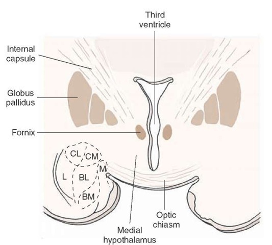
The amygdala is a prominent structure that lies deep within the temporal lobe at a level immediately rostral to the anterior limit of the hippocampal formation (Figs. 13-2 and 13-13). It consists of a lateral, central, basal, and medial nucleus (Fig. 13-16).One of the primary functions of the amygdala is to modulate processes normally associated with the hypothalamus and midbrain periaqueductal gray (PAG) matter. Examples of these functions include rage and aggression, flight and other affective processes, feeding behavior, endocrine and hormonal activity, sexual behavior, and autonomic control.
Modulation of these processes is achieved by virtue of major inputs from the amygdala to the hypothalamus and midbrain PAG. The most significant pathway of the amygdala is the stria terminalis. It arises from the medial amygdala and passes caudally and dorsally around the posterior thalamus as it follows the tail of the caudate nucleus to its subcallosal position adjoining the lateral ventricle (Fig. 13-13). The fibers then continue to follow the tail of the caudate nucleus in a rostral direction in which the strial fibers come to occupy a position just ventromedial to that of the caudate nucleus. At the level of the anterior commissure, the strial fibers descend in a ventromedial direction through the bed nucleus of the stria terminalis into the medial hypothalamus.
FIGURE 13-16 Cross-sectional diagram taken through the amygdala of a cat, illustrating the principal nuclei of the amygdala. BL = basolateral nucleus; BM = basomedial nucleus; CL = centrolateral division of the central nucleus; CM = centromedial division of the central nucleus; L = lateral nucleus of amygdala; M = medial nucleus of the amygdala.


