Cerebellar Cortex
Histology
The histological appearance of the gray matter is virtually identical throughout the entire cerebellar cortex. The gray matter forms folds (folia) that are arranged transversely within the long axis of the hemisphere (Figs. 21-8 and 21-11). Deep to the gray matter of each fold lies a massive region of white matter, which contains afferent and efferent axons of the cerebellar cortex.
The arrangement of the cells and fibers within the cer-ebellar cortex has important functional utility. For example, individual axons within the cerebellar cortex can affect many cells over wide areas of the cortex. Such an arrangement provides the basis by which the cerebellum influences wide regions of the body musculature at a given time. Another function of the cerebellum is to provide precise signals to distant structures in the form of excitatory-inhibitory sequences. Accordingly, the neurons within the cerebellar cortex must be arranged in such a manner that these signals can be appropriately transmitted to distal structures.
The cerebellar cortex consists of three cell layers (Fig. 21-8). The innermost layer is called the granular cell layer. The middle layer is the Purkinje cell layer, and the most superficial layer is called the molecular layer, which is almost devoid of cells. The anatomy and functions of the cells situated in these layers are described in the following sections.
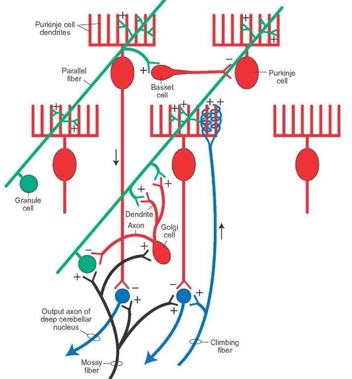
FIGURE 21-11 This figure shows some of the principal connections of the cerebellar cortex similar to those shown in Figure 21-10. This figure further illustrates how parallel fiber activation can lead to a beam of excitation within the plane of the parallel fiber and the Purkinje cell neurons with which they make synaptic contact and inhibition along adjacent beams of Purkinje cell neurons. The parallel fiber excites basket cells, which are inhibitory and make synaptic contact with Purkinje cells located in adjacent planes. This provides the basis for inhibition along the beam of Purkinje cells adjacent to the beam of excitation of Purkinje cells induced from the parallel fiber. The diagram also illustrates the relationship of the Golgi cell in inhibiting granule cell activity, as well as the inputs into the deep cerebellar nucleus, which constitutes the output neuron of the cerebellum. Such inputs include excitation from mossy and climbing fiber collaterals and inhibition from the Purkinje cells. Mossy fibers also excite granule cells, and climbing fibers excite Purkinje cells.
Granular Cell Layer
The primary cell found in the granular cell layer is the granule cell. This cell type contains three to five dendrites, which are arranged in a claw-like appearance to receive mossy fiber cerebellar afferents. The axon of the granule cell ascends toward the surface of the cortex and, as it enters the molecular layer, it ends by bifurcating and forming axons that pass in opposite directions within the molecular layer parallel to the surface of the cortex. These fibers are called parallel fibers. By passing along the long axis of the folium, the parallel fibers run in a direction perpendicular to the dendrites of Purkinje cells. Accordingly, this arrangement allows individual parallel fibers to make synaptic contact with many Purkinje cell dendrites. Parallel fibers serve to excite Purkinje cells, and these actions are believed to be mediated by glutamate. Furthermore, collaterals of parallel fibers make synaptic contact with dendrites of Golgi cells.
The other cell type found in the granular cell layer of the cerebellar cortex is the Golgi cell. It is an inhibitory, Y-aminobutyric acid (GABA)-ergic neuron. The cell body is typically found near the border of the Purkinje cell layer. In addition to receiving afferents from parallel fibers, the Golgi cell axon is short and makes synapse with dendrites of granule cells. Dendrites of Golgi cells also make synaptic contact with axon terminals of mossy fibers (Fig. 21-10).
Purkinje Cell Layer
The Purkinje cell layer contains one cell type, the Purkinje cell, and is, consequently, one cell in thickness. The cell body is flask-shaped, with the neck ascending vertically toward the molecular layer. Dendrites emerge from the neck, and thick dendritic trees are directed into the molecular layer (Fig. 21-8). The axon emerges from the bottom of the cell body and descends through the granular cell layer and white matter and terminates in one of the deep cerebellar nuclei. Purkinje cells located in the vermal region project to the fas-tigial nucleus, Purkinje cells located in the hemispheres project to the dentate nucleus, and Purkinje cells located in the paravermal region project to the interposed nuclei. Purkinje cell axons from the anterior lobe also project to the lateral vestibular nucleus. The Purkinje cell is inhibitory and uses GABA as a neurotransmitter.
Molecular Layer
The molecular layer contains the dendrites of Purkinje cells, parallel fibers (i.e., axons of granule cells), and two other cell types referred to as basket cells. The basket cells receive inputs from parallel fibers and make inhibitory axosomatic contacts with Purkinje cells (Fig. 21-10).
Functional Properties of the Cerebellar Cortex
As shown in Figure 21-10, stimulation of either the mossy or climbing fibers will result in activation of their target neurons. Consider first the flow of information through the cerebellar cortex after stimulation of the mossy fiber system. Mossy fibers excite granule cells, which, in turn, excite Purkinje cells. Within this circuit lies the Golgi cell, which can be activated in two ways. The first involves the parallel fiber, which makes synaptic contact with the dendrites of the Golgi cell. Alternatively, the mossy fiber can directly contact the dendrites of the Golgi cell. Golgi cell activation results in inhibition of the granule cell. Thus, inhibition of the granule cell can come about by either: (1) feed-forward inhibition (mossy fiber ^ [+] Golgi cell ^ [-] granule cell), or (2) feedback inhibition (mossy fiber ^ [+] granule cell ^ [+] Golgi cell ^ [-] granule cell). The net effect of this component of the overall circuit is that the granule cell discharge is limited by the coactivation of the Golgi cell following stimulation of the mossy fiber. In essence, this cerebellar circuit displays an "on-off" feature. Specifically, the mossy fiber turns on the granule cell, which then turns on the Purkinje cell. The granule cell is then turned off by the action of the Golgi cell. Because the granule cell is now turned off, the Purkinje cell and the Golgi cell are also turned off (called disfacilitation of the Purkinje and Golgi cells) because action potentials at this time are not conducted along the parallel fiber.
Concerning other elements of the cerebellar circuit, we have just indicated that parallel fiber activation, which is limited by the action of the Golgi cell, results in direct excitation of Purkinje cells that lie in a row within a given plane of the parallel fiber. The parallel fiber also can activate basket and stellate cells, which are inhibitory neurons. However, these neurons do not inhibit Purkinje cells that lie in the plane of excitation of the activated parallel fiber. Instead, these interneurons typically inhibit Purkinje cells that lie in an adjacent plane to that of the Purkinje cell excited by the parallel fiber (Fig. 21-11). Therefore, the overall process produces a beam of "excitation-inhibition" along a given plane of Purkinje cells, with a consequent inhibition of rows of Purkinje cells lying in adjacent planes. The net effect of this process is to sharpen the signals transmitted by the beam of Purkinje cells excited by the parallel fiber.
The action of the climbing fiber on the Purkinje cell is direct and involves no interneurons. The climbing fiber affects the discharge properties of Purkinje cells differently than do mossy fibers. Because a climbing fiber makes many synaptic contacts with a Purkinje cell, the effect of the climbing fiber is to produce a powerful depolarization of the Purkinje cell. Voltage-sensitive Ca2+ (calcium) channels propagate the ensuing action potentials in the Purkinje cell axon. As a result of the activation of the climbing fibers, the sensitivity of the Purkinje cell to other types of inputs becomes altered. Specifically, parallel fibers become less effective in causing excitation in the Purkinje cell for a significant period of time (1-3 hours). These long-term changes are most likely the result of initiation of second-messenger activity in the Purkinje cell.
It should be pointed out that the only way that signals can leave the cerebellar cortex is through the Purkinje cell axon, which is inhibitory to the deep cerebellar nucleus. How, then, does information get out of the cerebellar cortex? In answer to this question, recall that both mossy and climbing fibers send collaterals to the deep cerebellar nuclei. Therefore, mossy or climbing fiber activation results in an immediate excitation of the deep cerebellar nucleus. This excitation is then followed by inhibition mediated by the Purkinje cell. Because the duration of excitation of the parallel fiber is limited by the action of the Golgi cell, the excitatory input to the Purkinje cell from the parallel fiber is also of short duration. When that excitation ceases, there is a consequent increase in excitation in the deep cerebellar nucleus because the Purkinje cell is no longer excited (to produce inhibition on the deep cerebellar nucleus). This release of inhibition is referred to as disinhibition. Thus, the signals transmitted by the deep cerebellar nucleus to its target neurons in the brainstem or thalamus after mossy or climbing fiber stimulation would consist of initial excitation, followed by inhibition, which is then followed by disinhibition. This pattern of discharge then becomes the coded message transmitted from the cerebellum to distal targets as a part of an overall feedback response pattern. It should be pointed out that, although the output of the cerebellar cortex is inhibitory, the output of the cerebellum as a whole is excitatory. Inasmuch as the actions of the deep cerebellar nuclei on their target neurons are excitatory, these coded messages are further transmitted faithfully from the deep cerebellar nuclei to their target structures in the brainstem and thalamus and, ultimately, to the spinal cord and motor cortex.
Efferent Projections of the Cerebellar Cortex: The Feedback Circuitry
As noted earlier in this topic, the functional anatomical organization of the cerebellum may be divided into three categories: the vestibulocerebellum (i.e., flocculonodular lobe), spinocerebellum (anterior lobe), and cerebrocere-bellum (lateral hemispheres mainly of posterior lobe). The vestibulocerebellum is concerned with the status of the head region (in particular, control of the eyes and position of the head). The spinocerebellum is concerned with the status of the axial musculature (muscle tone) as well as its degree of flexion or extension at any given point in time. The cerebrocerebellum is concerned with the planning, organizational, and coordinating aspects of motor responses.
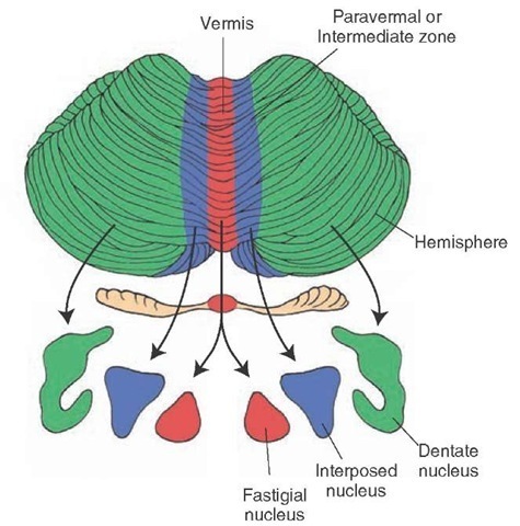
The cerebellar cortex transmits its signals to the deep cerebellar nuclei via the Purkinje cell axon, which is inhibitory. The inhibitory input from the Purkinje cell (which itself undergoes modification) thus alters the initial signal that the deep cerebellar nucleus receives from the mossy or climbing fiber and serves to sculpt the coded message transmitted to distant structures. The key point to remember is that the organization of the Purkinje cell projections to the deep cerebellar nuclei are topographically arranged. Purkinje cell axons associated with both the vestibulocerebellum and spinocerebellum project to the fastigial nucleus, whereas Purkinje cell axons associated with the cerebrocerebellum project to the interposed and dentate nuclei (Fig. 21-12). These relationships are central to our understanding of the functions of the cerebellum and disease states associated with different regions of the cerebellum.
FIGURE 21-12 Medial to lateral zonal organization of the cerebellar cortex with respect to its outputs to the deep cerebellar nuclei. The vermal region and its projection target in the fastigial nuclei are shown in red, the paravermal region and its projection target in the interposed nuclei are shown in blue, and the region of the hemisphere and its projection target in the dentate nuclei are shown in green.
Efferent Connections of the Vestibulocerebellum and Spinocerebellum
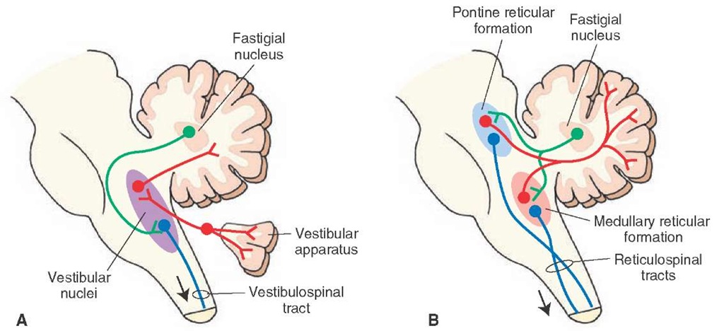
The fastigial nucleus, which is related to functions of both the vestibulocerebellum and spinocerebellum, has two primary targets within the brainstem to which its axons project: the vestibular nuclei and reticular formation. The fibers project topographically and bilaterally to vestibular nuclei and contralaterally to the parts of the reticular formation of the pons and medulla near the origins of the reticulospinal pathways (Fig. 21-13).
These connections thus establish two of the feedback circuits governing the axial musculature. One circuit involves the vestibular nuclei with the cerebellum, and the other involves the reticular formation with the cerebellum. The vestibular feedback circuit can be summarized as follows: vestibular input from the vestibular apparatus ^ vestibular nuclei (or primary vestibular fibers direct to) ^ flocculonodular lobe and posterior vermis ^ fastigial nucleus ^ vestibular nuclei ^ (to extensor motor neurons of spinal cord and cranial nerve nuclei III, IV, VI).
Thus, the vestibular-cerebellar feedback pathway enables the cerebellum to regulate the activity of neurons in the medial and lateral vestibular nucleus that control motor neuron activity within the spinal cord and brain-stem. Concerning functions of the vestibulocerebellum and the medial vestibular nucleus, the feedback connections to the vestibular nuclei allow for cerebellar control over the position of the eyes via the vestibular inputs into the medial longitudinal fasciculus and over the position of the head via its connections with the medial vestibular nucleus and the medial vestibulospinal tract (which influences the position of the head). In this manner, changes in postural position and head position result in activation of the vestibulocerebellum, which, in turn, transmits signals back to the vestibular nuclei that can modify the position of the head and eyes. Likewise, inputs into the lateral ves-tibular nucleus modulates the activity of the lateral vestibulospinal tract, which mediates control over limb extensor muscles and axial muscles, including motor tone of these muscles.
FIGURE 21-13 Diagrams illustrate the output relationships from the fastigial nucleus of cerebellum with (A) vestibular nuclei and spinal cord and (B) reticular nuclei and spinal cord. Inputs to the cerebellar cortex from the reticular formation and vestibular nuclei are shown in red, outputs of the fastigial nuclei of the cerebellum to the reticular formation and vestibular nuclei are shown in green, and the outputs of the reticular formation and vestibular nuclei (i.e., reticulospinal and vestibulospinal tracts) to the spinal cord are shown in blue.
FIGURE 21-14 Diagram depicts the outflow pathways from the cerebellar cortex to the cerebral cortex and red nucleus. The linkage between the cerebellum and cerebral cortex involves a disynaptic pathway—an initial projection from the dentate nucleus to the ventrolateral thalamic nucleus and a second projection from the thalamus to the motor and premotor cortices (both shown in red). The pathway from the cerebellum to the red nucleus involves a projection from the interposed nuclei to the red nucleus (shown in green). Both of these outputs from the cerebellum to the contralateral thala-mus and red nucleus are mediated through the superior cerebellar peduncle. The corticospinal and rubrospinal pathways are indicated in blue.
With respect to the spinocerebellum, the same general circuitry is involved through the feedback connection to the lateral vestibular nucleus. In particular, the inhibitory inputs to the lateral vestibular nucleus from Purkinje cell axons in the anterior lobe play an important role in extensor motor tone and general control over axial muscles and limb extensors. The control over muscle tone by the spinocerebellum is further mediated via the connections of the fastigial nucleus with the reticular formation of the pons and medulla. This circuitry, the reticular-cerebellar circuit, can be summarized as follows: nuclei of the reticu-lar formation of the medulla and pons ^ anterior and posterior lobes (vermal and paravermal regions) ^ fastigial nucleus ^ nuclei of reticular formation of the pons and medulla (that project to extensor motor neurons of spinal cord).
This feedback circuit shares similarities with that of the vestibular-cerebellar feedback circuit in that both the reticular formation and lateral vestibular nucleus act on extensor motor neurons of the spinal cord and, thus, serve to regulate muscle tone and one’s ability to stand erect. Thus, when taken collectively, the cerebellar cortical relationships with these brainstem nuclei and, ultimately, with the spinal cord provide the feedback mechanisms by which the spinocerebellum can influence extensor motor processes within the spinal cord (Fig. 21-13).


