The cerebellum is concerned with at least three major functions. The first function is an association with movements that are properly grouped for the performance of selective responses that require specific adjustments. This is also referred to as synergy of movement. The second function includes the maintenance of upright posture with respect to one’s position in space. The third function concerns the maintenance of the tension or firmness (i.e., tone) of the muscle.
If one were to analyze what is required neurophysiolog-ically to complete even the simplest movements, such as walking or lifting a fork to one’s mouth, it would be apparent that they are, indeed, complex acts. To be able to complete either of these responses, the following elements are required: (1) contraction of a given muscle group or groups of muscles, (2) simultaneous relaxation of antagonist set(s) of muscles, (3) specific level of muscle contraction for a precise duration of time, and (4) the appropriate sequencing of contraction and relaxation of the muscle groups required for the movement in question. The numbers of muscle fibers activated determines the extent of the muscle contraction. In turn, the numbers of muscle fibers that contract at a given time (i.e., the force or strength of contraction) are a function of the numbers of alpha motor neurons that are activated. The duration of contraction is determined, to a large extent, by the duration of activation of the nerve fibers that innervate the muscles required for the specific act.
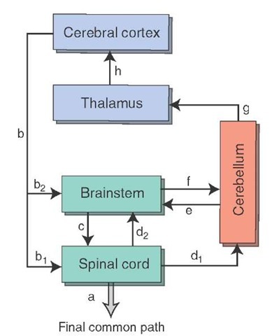
For precise and effective execution of purposeful movements as well as the presence of appropriate posture in association with standing and with movement, a region of the brain must be able to integrate and organize the sequence of events associated with the response. Such a region should be able to both receive inputs from all the regions of the central nervous system (CNS) associated with motor function and, consequently, send "feedback" responses to these regions. Thus, such a region must function, as a computer does, to integrate sensory and motor signals and, consequently, it must have the necessary computer-like or integrative properties for analysis of the afferent signals and possess the reciprocal connections to form a series of "feedback" pathways to its afferent sources. The region of the brain possessing all of these characteristics is the cerebellum. It receives inputs from all regions of the CNS associated with motor functions and sensory regions mediating signals about the status of a given muscle or groups of muscles (Fig. 21-1). It also has the capacity to send back messages to each of these regions. Moreover, the cerebellum possesses the machinery for integrating each of these afferent signals. The remainder of this topic is designed to document how the cerebellum carries out these functions.
GROsS ORgANIZATION of the CEReBELLUM
A description of the gross anatomical characteristics of the cerebellum and the student is referred to that topic for details concerning morphological characteristics.
FIGURE 21-1 Relationships of the cerebellum with other regions of the central nervous system. In this scheme, the final common path (a) for the expression of motor responses is mediated through the spinal cord. These motor responses require inputs to the cerebellum from the cerebral cortex (b) via the brainstem (b2 and f) as well as through internal reflex mechanisms contained within the spinal cord (more directly from b, and d, or indirectly from b,, d2, and f). The appropriate "feedback" from the cerebellum to the structures that provide input to it include outputs to the spinal cord through brainstem structures (e + c) and to the cerebral cortex (g + h).
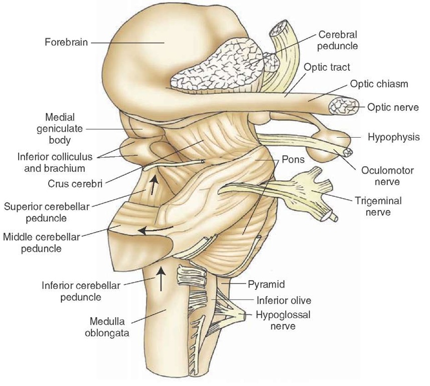
This material is briefly summarized here. The cerebellum is attached to the brainstem by three peduncles. The inferior and middle cerebellar peduncles are mainly cerebellar afferents, whereas the superior cere-bellar peduncle contains mainly cerebellar efferent fibers. The inferior cerebellar peduncle emerges from the dorsola-teral medulla and enters the cerebellum on its inferior aspect. The middle cerebellar peduncle emerges from the lateral aspect of the pons and enters the cerebellum from a lateral trajectory. The superior cerebellar peduncle emerges from the superior and medial aspects of the cerebellum and enters the brainstem at the level of the upper pons (Fig. 21-2).
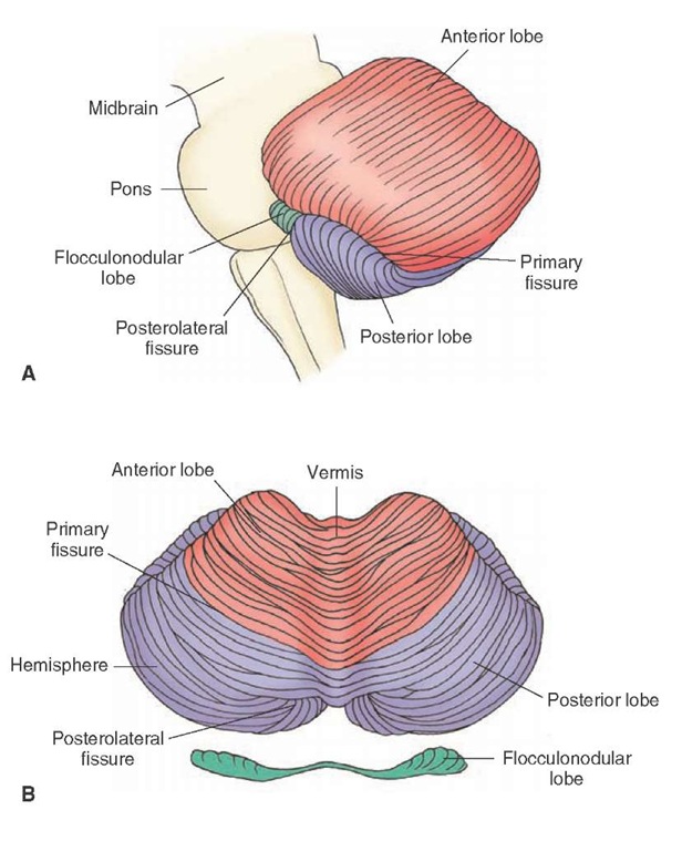
The cerebellar cortex consists of three lobes. Each lobe consists of two bilaterally symmetrical hemispheres separated from each other by a midline structure called a vermis (Fig. 21-3). The lobes are identified as the anterior, posterior, and flocculonodular lobes. The anterior lobe is separated from the posterior lobe by the primary fissure, and the posterior lobe is separated from the flocculonodu-lar lobe by a posterolateral fissure. Phylogenetically, the flocculonodular lobe is the most primitive. It receives major inputs from the vestibular system and is, accordingly, referred to as the vestibulocerebellum. The anterior lobe and adjoining parts of the vermal region of the posterior lobe receive major spinal cord inputs. Therefore, this is referred to as the spinocerebellum. Because the anterior lobe evolved at an earlier time than the posterior lobe, it is sometimes also called the paleocerebellum. The largest and most recently (from a phylogenetic viewpoint) evolved region of the cerebellum is the posterior lobe.
FIGURE 21-2 Lateral view of the brainstem indicating the relationships of the cerebellar peduncles to the brainstem and cerebellum. Arrows denote the general direction of information flow with respect to the cerebellum. The inferior and middle cerebellar peduncles contain mostly cerebellar afferent fibers, while the superior cerebellar peduncle contains mainly cerebellar efferent fibers.
FIGURE 21-3 The cerebellum. Note the general fissures and divisions of the vermis as illustrated in (A) sagittal and (B) dorsal representations.
It is linked most closely with the cerebral cortex. Accordingly, it is also called the neocerebellum. Thus, one can see that the cerebellar cortex can be divided into three different functional regions. Their significance will be discussed later.
Deep to the cerebellar cortex is a large expanse of white matter that separates the cortex from the underlying deep cerebellar nuclei. There are three deep cerebellar nuclei represented bilaterally. The medial structure is called the fastigial nucleus. The lateral structure is the dentate nucleus, and the nuclei that lie between the dentate and fastigial nuclei are called the interposed nuclei (see Fig. 11-8). The interposed nuclei consist of two relatively small nuclei; the medial structure is called the globose nucleus, and the lateral one is called the emboliform nucleus. Each of these deep cerebellar nuclei mediates different functions, which relate to their input-output properties. The functions of these nuclei are discussed later in this topic.
Afferent Connections of the Cerebellum
The cerebellar cortex receives inputs from regions of the CNS associated with the regulation of motor functions as well as direct and indirect sensory inputs associated with the status of i ndividual muscles, groups of muscles, and other inputs that include tactile impulses, cutaneous affer-ents, and high-threshold joint afferents. The following sections elaborate on the sources, distribution, and functional nature of the afferent supply to the cerebellar cortex.
Spinal Cord (Spinocerebellum)
The inputs from the spinal cord provide the cerebellum with essential information (i.e., the status and position of individual as well as groups of muscles) with which it can control both muscle tone and the execution of movements. The pathways most critical for supplying such inputs to the cerebellum include the dorsal spinocerebellar tract, ventral spinocerebellar tract, cuneocerebellar tract, and the rostral spinocerebellar tract (the latter identified in the cat). The projections of these pathways to the cerebellum are somatotopically organized and are directed primarily to the vermis and regions around the vermis (paravermal region) of the anterior lobe and adjacent portions of the posterior lobe. Therefore, these receiving areas are referred to as the spinocerebellum.
Dorsal (Posterior) Spinocerebellar Tract
Therefore, only the key features of these tracts will be briefly summarized at this time.
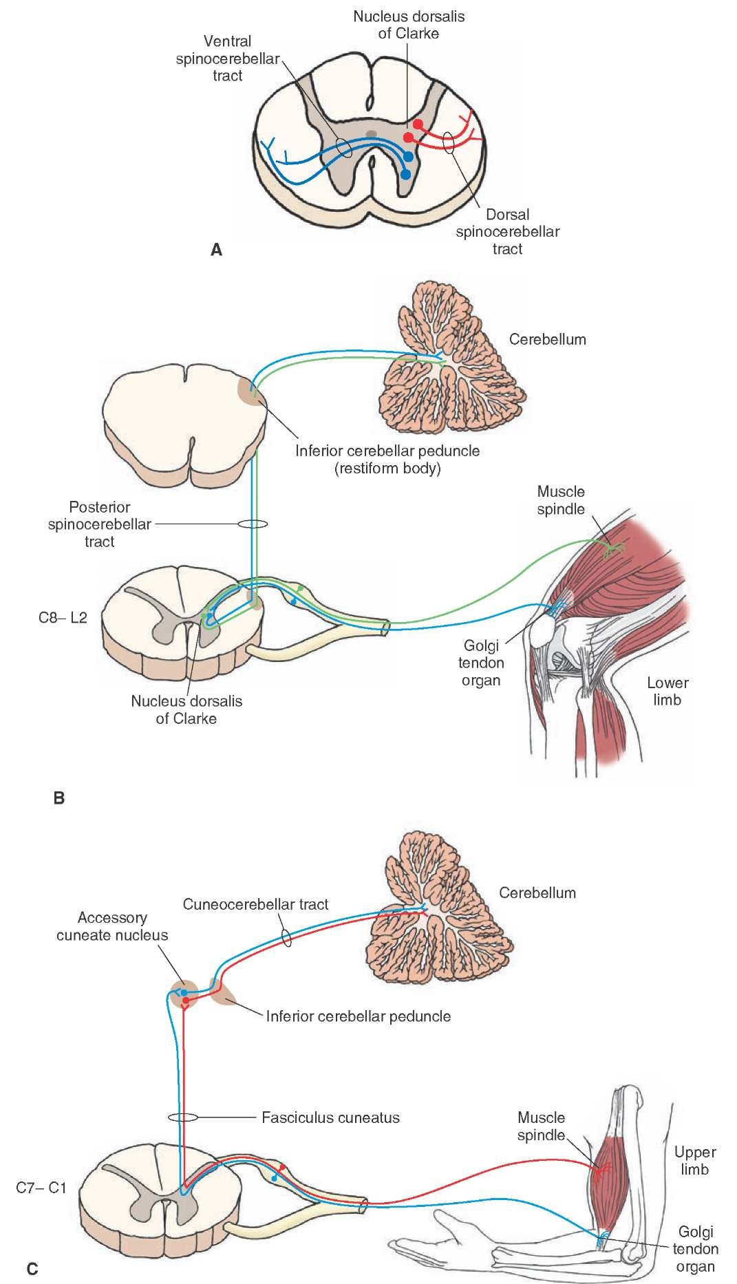
The dorsal spinocerebellar tract, which conveys signals mainly from muscle spindles and Golgi tendon organs concerning the status of individual muscles to the cerebellar cortex from the lower limbs, passes through the inferior cerebellar peduncle and terminates mainly in the medial part of the ipsilateral anterior lobe and adjacent portions of the posterior lobe (Fig. 21-4, A and B).
Ventral (Anterior) Spinocerebellar Tract
The synergy of movement conveys signals from the Golgi tendon organ (i.e., detecting whole limb movement) and flexor reflex afferents1 through the superior cerebellar peduncle to the anterior lobe of the cerebellum close to the regions where dorsal spinocerebellar fibers terminate (see Figs. 21-4A and 9-9).
Cuneocerebellar Tract
The cuneocerebellar tract, the upper limb equivalent of the dorsal spinocerebellar tract, conveys inputs from muscle spindles of the upper limb to the upper limb regions of the anterior lobe and adjoining regions of the posterior lobe (Fig. 21-4C). This pathway thus provides the spinoc-erebellum with inputs concerning mainly the status of individual muscles of the upper limb.
Rostral Spinocerebellar Tract
An upper limb equivalent of the ventral spinocerebellar tract not yet identified in humans, the rostral spinocere-bellar tract arises from the cervical cord and supplies the anterior lobe of the cerebellum, conveying whole limb movement (from the Golgi tendon organ) to the anterior lobe from the upper limb.
Brainstem
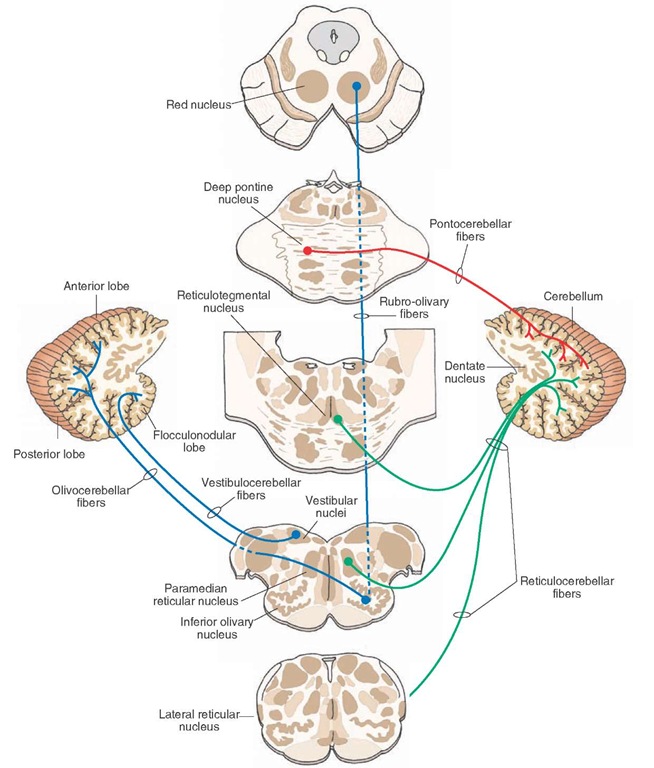
A number of nuclei in the brainstem contribute inputs to the cerebellum. These structures include the inferior olivary nucleus, vestibular nuclei, several groups of nuclei within the reticular formation, and deep (or ventral) pon-tine nuclei. The nature and functions of these cerebellar afferent fibers from each of these brainstem nuclei vary and are considered in the following sections.
Inferior Olivary Nucleus
The largest component of the inferior cerebellar peduncle is associated with fibers arising from the inferior olivary nucleus. The inferior olivary nucleus receives two different kinds of inputs. One type of input is from the spinal cord and the second is from the cerebral cortex.
Fibers from the spinal cord convey information from cutaneous afferents, joint afferents, and muscle spindles to the inferior olivary nucleus. From this structure, axons pass to the contralateral cerebellar cortex via the inferior cere-bellar peduncle, terminating somatotopically within the anterior and posterior lobes of the cerebellar cortex (Fig. 21-5).
The second kind of input relates to the sensorimotor cortex and includes motor fibers that descend from the cerebral cortex directly and indirectly via the red nucleus.
FIGURE 21-4 Origin and distribution of spinocerebellar and cuneocerebellar tracts. (A) The origins of the dorsal and ventral spinocerebellar tracts within the spinal cord. (B) Origin, course, and distribution of the posterior spinocerebellar tract and (C) cuneocerebellar tract. C = cervical.
FIGURE 21-5 The afferent pathways to the cerebellar cortex from the brainstem. These include pathways arising from the red nucleus (shown in blue), deep pontine nuclei (shown in red ), vestibular nuclei, and reticular formation.
In humans, the rubro-olivary fibers are much more extensive than in other species of animals. In this manner, the inferior olivary nucleus appears to transmit an integrated signal from the spinal cord and cerebral cortex to the cer-ebellar hemispheres via the climbing fiber system.
