The midbrain lies between the pons and the forebrain (Figs. 12-1 and 12-2). When viewed in cross section at either rostral or caudal levels of the midbrain, it is customary to divide this region of the brainstem into three anatomically distinct components (Fig. 12-3). (1) The most dorsal part is called the tectum. It is sometimes referred to as the corpora quadrigemina, which means "four bodies" (i.e., the superior and inferior colliculi appear as four rounded bodies when looking down on the midbrain). (2) The ventral aspect is referred to as the crus cerebri. It includes massive bundles of fibers that pass from the cerebral cortex to the brainstem and spinal cord. (3) The central part of the midbrain is called the tegmentum, which is continuous with the tegmentum of the pons. A cell group called the substantia nigra separates the tegmentum from the crus cerebri. The cerebral aqueduct, which is surrounded by gray matter called the periaqueductal gray (PAG), separates the tegmentum from the tectum (Fig 12-3).Some authors divide the midbrain into two parts: a tectal region, as described earlier, and a cerebral peduncle, which includes both the crus cerebri (located ventrally) and the tegmentum (located dorsally).
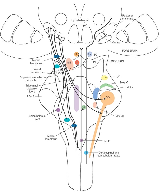
FIGURE 12-1 The loci of the major cell groups and fiber tracts at the level of the midbrain. IC = inferior colliculus; LC = locus ceruleus; MLF = medial longitudinal fasciculus; Mes V = mesencephalic nucleus of cranial nerve (CN) V; MO V = motor nucleus of CN V; MO VII = motor nucleus of CN VII; RN = red nucleus; SC = superior colliculus; SN = substantia nigra; VI = nucleus of CN VI; IV = nucleus of CN IV; III = nucleus of CN III.
FIGURE 12-2 Midsagittal section of the brainstem depicting the midbrain and the levels at which the cross-sectional diagrams were taken, as indicated in Figures 12-3 and 12-4.
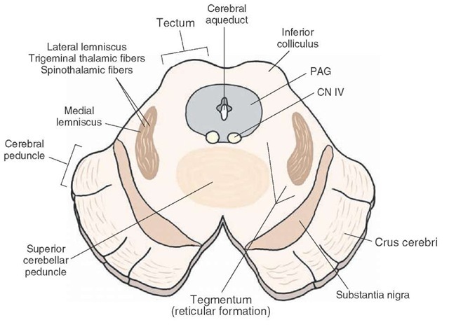
FIGURE 12-3 Cross-sectional diagram in which the principal structures of the midbrain at the level of the inferior colliculus are depicted. Note the basic divisions of the midbrain into the tectum, tegmentum, and crus cerebri. PAG = periaqueductal gray; CN = cranial nerve.
The midbrain contains two cranial nerves. The trochlear nerve (cranial nerve [CN] IV) emerges on the dorsal aspect of the caudal midbrain just caudal to the inferior colliculus (Figs. 12-3 and 12-4). The oculomotor nerve (CN III) emerges on the ventromedial aspect of the midbrain at a position just medial to the crus cerebri (Fig. 12-5).
Internal Organization of The Midbrain
Because of the arrangement of the principal cell groups and fiber bundles of the midbrain, it is convenient to examine the organization of these structures at two levels. The caudal half includes the level of the inferior collicu-lus, and the rostral half includes the level of the superior colliculus.
Level of the Inferior Colliculus
Tectum
In the caudal half of the midbrain, the tectal region contains the inferior colliculus (Figs. 12-1, 12-3, and 12-4). The inferior colliculus consists of a large nuclear structure that represents an important relay component of the auditory pathway. It receives ascending inputs from auditory relay nuclei of the medulla and pons.
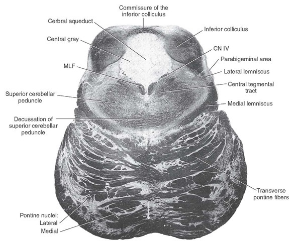
FIGURE 12-4 Photograph of a cross section taken at the level of the inferior colliculus (Weigert stain). MLF = medial longitudinal fasciculus; N.IV = cranial nerve IV.
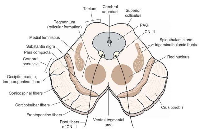
FIGURE 12-5 Cross-sectional diagram in which the principal structures of the midbrain at the level of the superior colliculus are depicted. CN = cranial nerve; PAG = periaqueductal gray matter.
FIGURE 12-6 Photograph of a cross section taken at the level of the superior colliculus (Weigert stain). N. III = cranial nerve III.
These groups of neurons supply the inferior colliculus via a pathway called the lateral lemniscus, which ascends in the lateral aspect of the pons and caudal midbrain (Fig. 12-3). The inferior colliculus, in turn, projects its axons through a fiber bundle called the brachium of the inferior colliculus (to the medial geniculate nucleus of the thalamus) (see Fig. 12-6, which does not include the medial geniculate nucleus because it lies at a more rostral position).
Tegmentum (Including the Periaqueductal Gray Matter)
The tegmentum includes a variety of cell groups and fiber tracts. The PAG, a transitional region between the tectum and tegmentum, is composed mainly of tightly packed cells that surround the cerebral aqueduct (Figs. 12-3 and 12-5). Although not typically visible through normal stained material, axons arising from both the PAG as well as from regions of the forebrain descend through different levels of the PAG to terminate either within the PAG or at lower regions of the brainstem. The PAG contains high concentrations of the neurotransmitter peptide, enkephalin.The PAG plays important roles in the regulation of autonomic functions and affective and emotional processes and in the modulation of pain impulses.
Just beneath the PAG lies the nucleus of the trochlear nerve (a general somatic efferent [GSE] nucleus). Its axons pass dorsally and caudally until they exit the brain on the contralateral side at the caudal aspect of the inferior col-liculus.These axons then innervate the superior oblique muscle, which moves the eye downward when it is displaced medially. Damage to this cranial nerve is manifest, in particular, in the form of diplopia when the patient tries to look downward such as when attempting to walk down a flight of stairs. Beneath the nucleus of the trochlear nerve lies the medial longitudinal fasciculus; this serves as an afferent source to the tro-chlear nucleus (Fig. 12-4).
The lateral aspect of the tegmentum contains a number of important ascending sensory pathways. These pathways include the lateral lemniscus, which provides auditory inputs to the inferior colliculus from other auditory relay nuclei of the upper medulla, and the spinotha-lamic, trigeminothalamic, and medial lemniscal pathways (Figs. 12-1 and 12-3).
Between the medial and lateral aspects of the tegmen-tum lies the central tegmental area. The central tegmental area contains the reticular formation, which extends ros-trally from the medulla as described previously (Fig. 12-4). At this level of the brainstem, the reticular formation contains a variety of nuclei and fiber tracts. Along its medial edge can be found raphe nuclei, which include serotonin-containing neurons that project to the forebrain and to lower regions of the brainstem. Nuclei of this region of the tegmentum also contribute to the regulation of somatomo-tor, autonomic, and other visceral processes.
Embedded within the tegmentum at this level of the mid-brain is the decussation of the superior cerebellar peduncle (Fig. 12-4). The superior cerebellar peduncle originates from the dentate and interposed nuclei of the cerebellum and passes into the brainstem at the level of the upper pons. At the level of the inferior colliculus, these fibers decussate as they continue to ascend to the red nucleus and ventrolateral nucleus of the thalamus, where they terminate (Fig. 12-5).
Crus Cerebri
The ventral aspect of the brainstem at this level contains the crus cerebri (Fig. 12-5). As described earlier, the crus cerebri contains massive bundles of descending axons from the cerebral cortex that terminate either within the brainstem (i.e., corticobulbar fibers) or within the spinal cord (i.e., corticospinal fibers). This peduncle is clearly organized in the following manner. Fibers in the lateral fifth arise from the parietal, occipital, and temporal neo-cortices and terminate upon deep pontine nuclei. Fibers contained in the medial fifth also terminate upon deep pontine nuclei, but they arise from the frontal lobe. Fibers contained within the medial three fifths constitute the corticobulbar and corticospinal tracts (Figs. 12-1 and 12-5). The organization of these fibers is such that fibers associated with the head region are located medially (cor-ticobulbar fibers); whereas fibers associated with the upper limb, trunk, and lower limbs are located laterally.
Situated in a transitional position between the crus cer-ebri and the tegmentum is the substantia nigra. It contains two groups of cells; one group is located medially and is highly compacted (called the pars compacta), and one is located laterally, reticulated in appearance, and called the pars reticulata (Fig. 12-5). Each of these regions is important because of the neurotransmitters that they synthesize as well as their projection targets. Dopamine, for example, is associated with the pars compacta and is released onto neurons of the neostriatum. In contrast, neurons of the pars reticulata project to the thalamus, using gamma-aminobutyric acid (GABA) as a neurotransmitter. Clinically, it is known that loss of dopaminergic neurons in the substantia nigra results in a motor disorder called Parkinson’s disease.


