The terminology and essential features of the pulp chambers and pulp canals are considered before presenting the details of pulp chambers and canals using sectioned tooth specimens. Then a brief section on radiographic visualization of pulp chambers and canals is provided. After that a short section is presented on crown and root fractures. A final section considers the relationship of the teeth to the mandibular canal.
The use of the term pulp chamber, pulp cavity, or coronal pulp to designate that part of the crown normally filled with soft tissue varies with the anatomist; however, as with the terms root pulp and radicular pulp, and pulp canal and root canal, it is probably a matter of professional preference, because a good case can be made for each of the terms. These two sets of terms relating to crown and root are used as if they have the same meaning, with the recognition that arguably there may be contextualized differences.
Pulp, Chamber, and Canals
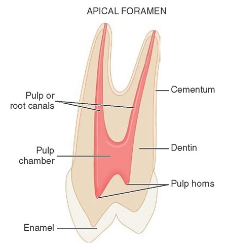
The crown and root portion of a tooth that contains the pulp tissues has been arbitrarily divided into the pulp chamber and the root or pulp canal (Figure 13-1). The complexities of these cavities cannot be fully appreciated without studying longitudinal and transverse sections of each of the representative types of teeth.
The dental pulp is the soft tissue component of the tooth. It occupies the internal cavities of the tooth (i.e., the pulp chamber and pulp canal). In general, the outline of the pulp tissue corresponds to the external outline form of the tooth (i.e., the outline form of the pulp chamber corresponds with the shape of the crown, whereas the outline form of the pulp canal corresponds with the shape of the roots of a tooth).
The dental pulp within these cavities originates from the mesenchyme and has been assigned a number of different functions: formative, nutritive, sensory, and defensive. The initial function of the dental pulp is the formation of dentin during the developmental period. The complex sensory system within the dental pulp controls the blood flow and is responsible for at least mediation of the sensation of pain. The formation of reparative dentin or secondary dentition (osteoid-like dentin) represents a defensive response to any form of irritation, whether it is mechanical, thermal, chemical, or bacterial. The reactive dentin is usually limited to the area of pulpal irritation. Separating reactive changes (response to injury) from purely aging-related changes may be difficult or impossible to do at this time.
Radiographs
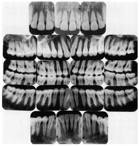
The use of radiographs or digital radiography for the diagnosis and treatment of pulpal disease requires that the morphological features of the pulp chambers and root canals, which are three-dimensional, be visualized when compressed into a "two-dimensional" radiographic image. Thus, radiographic views taken of the teeth from a facial orientation show a monoplane, buccolingual view of the hard tooth structures and radiolucent spaces for the pulp and canals (Figure 13-2). Mesiodistal aspects of longitudinal sections usually are seen only incidentally (e.g., on radiographs of malposed, rotated teeth). Thus, the radiographic anatomy of the pulp cavity from a mesial-distal aspect is not well known. Radiographic views of the pulp chambers and canals will be considered in more detail later.
FIGURE 13-1 Buccolingual section of a maxillary premolar.
SIZE OF THE PULP CAVITY
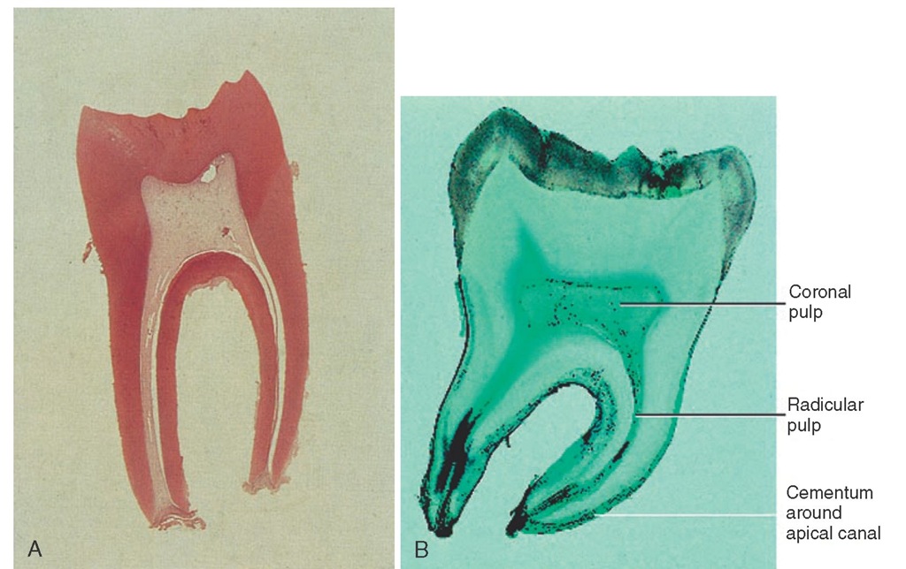
The size of the pulp chamber depends on the age of the tooth and its history of trauma. Secondary dentin is formed continuously throughout the life of the tooth as a normal process, as long as the vitality of the tooth is maintained. The formation of secondary dentin is not uniform, because the odontoblasts adjacent to the floor and roof of the pulp cavity produce greater quantities of secondary dentin than do the odontoblasts located adjacent to the walls of the pulp cavity.1 Therefore the size of the pulp cavity is much larger in a young individual than in an adult (Figure 13-3, A and B) and should be considered before extensive tooth reduction is accomplished, especially in a young person.
Various traumatic injuries occur that, if severe enough, will initiate a different type of dentin formation. Irritation-induced or reparative dentin may be formed in response to the carious process, abrasion, and attrition, as well as to operative procedures. This response is protective but may ultimately be detrimental in later years, because a finite amount of space is present within the pulp cavity. The size of the pulp cavity in a given tooth should be compared with that in the other teeth. If the calcification demonstrated is a localized phenomenon and is extensive, elective endodontic therapy is strongly suggested before any restorative procedure. Elective endodontics should be considered when extreme calcification is present in a tooth scheduled for complex restorative procedures.
Figure 13-2 Dental radiographic examination.
FIGURE 13-3 Comparison in size of the pulps of two intact lower first permanent molars at different ages. A, The young pulp chamber is large (magnification x8). B, The older pulp is greatly reduced in size (magnification x8).
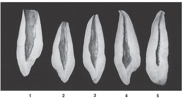
Figure 13-4 Maxillary canine. Labiolingual sections show various stages of development. 1, Crown complete; root partially completed with large pulp cavity, wide open at apical end. 2, Tooth almost complete, except for lack of constriction of apical foramen. 3, Canine of young individual with large pulp cavity and completed root tip. 4, Typical canine of adult, demonstrating constriction of the foramen. 5, Canine of an elderly individual with a constricted pulp chamber and canal; this specimen has lost its original crown form because of wear during function.
Foramen
The neurovascular bundle, which supplies the internal contents of the pulp cavity, enters through the apical foramen or foramina (see Figure 13-1). As the root begins to develop, the apical foramen is actually larger than the pulp chamber (Figure 13-4, 1), but it becomes more constricted at the completion of root formation (Figure 13-4, 2 through 5).
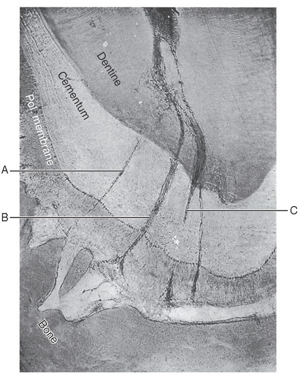
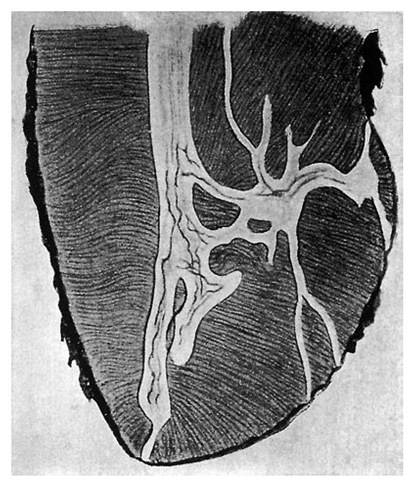
It is possible for any root of a tooth to have multiple apical foramina. If these openings are large enough, the space that leads to the main root canal is called a supplementary or lateral canal (Figure 13-5). If the root canal breaks up into multiple tiny canals, it is referred to as a delta system,2 because of its complexity (Figure 13-6).
Demarcation of Pulp Cavity and Canal
The cementoenamel junction (CEJ) is not quite at the level at which the root canal becomes the pulp chamber (see Figure 13-1). This demarcation is mainly macroscopically based but may be visualized by exploring the CEJ (see Figure 2-16) and noting the difference in density between the enamel and dentin at the mesial and distal tooth surfaces on radiographs. Enamel covers the external surface of the dentin, which makes up part of the pulp chamber, whereas cementum covers the entire external dentinal surface of the root canal space. The demarcation is simpler in multi-rooted teeth, because the pulp cavity within the root is the root canal and the remaining pulp cavity is the pulp chamber. Microscopically, the pulp within the chamber appears to be more cellular than the pulp found within the pulp of the root canal. The odontoblasts are cuboidal in the coronal pulp chamber but gradually flatten out as the apex is approached. The transition from the pulp chamber to the root canal is not sharply demarcated microscopically, and this demarcation is not sharply delineated macroscopically.
Figure 13-5 A section through the apex of a root showing multiple canals (A through C). Three canals are present within the dentin, but one canal divides at the cementum-dentin junction (C), which makes a total of four small canals that exit on the cemental surface of the tooth (Talbot).
Figure 13-6 Apical end of root showing one main canal and an adjacent delta system.
Pulp Horns
Projections or prolongations in the roof of the pulp chamber correspond to the various major cusps or lobes of the crown. The pulpal tissues that occupy these prolongations are called pulp horns (Figure 13-7). The prominence of the cusps or lobes corresponds with the development of the pulp horns. If the cusps or labial lobes are prominent (as in young individuals), one should expect to find equally prominent pulp horns underlying these structures (see Figure 13-8, B, 6). These projections become less prominent with time as a result of the formation of secondary dentin (see Figure 13-8, B, 1).
