Occlusal Contact Relations and Intercuspal Relations of the Teeth
The paths made by the supporting cusps of the maxillary and mandibular first molars in lateral and protrusive mandibular movements serve to illustrate the relationship of these cusps to the morphological features of these teeth (Figure 16-38).
Movements Away From Centric/ Eccentric Movements
Occlusal contact relations away from the intercuspal position (centric occlusion) involve all possible movements of the mandible within the envelope of border movements shown in Figures 15-13 and 15-14). These movements are generally referred to as lateral, lateral protrusive, protrusive, and retrusive movements. Lateral and lateral protrusive movements may be either to the right or to the left. Designations of lateral movement often do not include lateral protrusive movements, so that basic movements are reduced to right and left lateral movement, protrusive movement, and retru-sive movement.
Lateral Movements
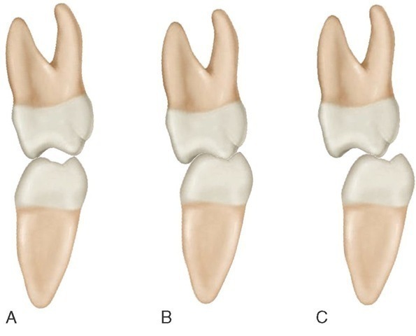
During the right lateral movement, the mandible is depressed, the dental arches are separated, and the jaw moves to the right and brings the teeth together at points to the right of the intercuspal position (centric occlusion) in right working (Figure 16-39, A). On the left side, called the nonworking side (or, for complete dentures, the balancing side), the teeth may or may not make contact (Figure 16-39, C). Condylar movement on the working side is termed a laterotrusive movement in the horizontal plane. The nonworking side condylar movement is a mediotrusive movement.
Tooth Guidance
Concepts of occlusion often describe idealized contact relations in lateral movements as shown in Figure 16-40.
Figure 16-39 Right side contact relations of first maxillary and mandibular molars: A, Right working side. B, Centric occlusion (intercuspal position). C, Nonworking side.
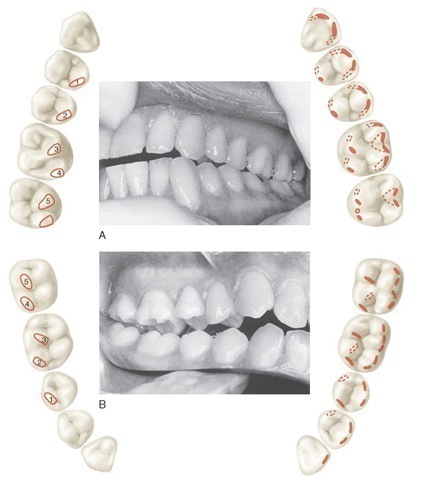
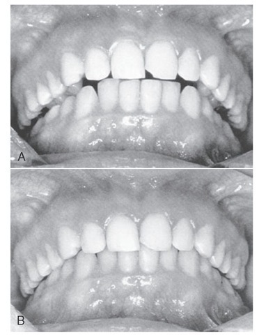
However, in the natural dentition, a variety of contact relations may be found, including group function, cuspid disocclusion only, or some combination of canine, premolar, and molar contacts in lateral movements. Group function refers to multiple contacts in lateral or eccentric mandibular movements (Figure 16-41, A) rather than simply canine (cuspid) guidance (Figure 16-41, B). Incisal guidance refers to contact of the anterior teeth during protrusive movements of the mandible. Condylar guidance and neuromuscular guidance are considered later.
PROTRUSIVE MOVEMENTS
During protrusive movements, the mandible is depressed and then moves forward, bringing the anterior teeth together at points most favorable for the incision of food. A retrusive movement follows protrusive movement to the intercuspal position.
OBTAINING CENTRIC RELATION
Guidance by the clinician of the jaw into centric relation (Figure 16-42, A and B) and then into initial contact with the teeth (centric relation contact), occurs usually with one or two (premature) contacts. Often the contacts are either on the oblique ridge of the maxillary molar and/or the mesial cusp ridge of the maxillary first premolar (Figure 16-43). In the centric relation position the condyle-disk assemblies are positioned on the anterior slope of the mandibular fossae (Figure 16-44). After initial contact of the teeth the patient can squeeze the teeth together and the mandible will slide forward or eccentrically, depending on the position of the interference (Figure 16-45). This movement from the premature contact in centric relation to centric occlusion (intercuspal position) is called a slide in centric. Centric occlusion and intercuspal position are both maximal intercuspation. If the occlusal interferences in centric relation contact are removed by selective grinding, the ability to close into maximum intercuspation without interference any place between centric relation and centric occlusion is called freedom in centric (see Figures 15-13 and 15-14).
RETRUSIVE MOVEMENTS
Retrusive movement from centric occlusion to retruded contact position in which the condyles are in the rearmost, uppermost position seems to occur in bruxism but infrequently in mastication and swallowing, except when centric occlusion and centric relation are coincident or when maximum intercuspation occurs at centric relation contact. The adverse neuromuscular responses that may occur to retrusion and closure of the jaw in the presence of occlusal interferences to maximum intercuspation in centric relation can be detected electromyographically and by the clinician. However, the jaw does not go to the retrusive position necessarily even after elimination of such occlusal interferences by occlusal adjustment. Although the occlusion (anterior guidance) may be reconstructed to guide the mandible into the retruded contact position and maximum intercuspation in centric relation, a movement from centric relation to the intercuspal position (slide in centric) may develop again. The significance of the return of a small slide from centric relation occlusion to the intercuspal position (centric relation to occlusion) has not been determined.45 The return of a slide occurs with orthodontics as well.46
Lateral Occlusal Relations
When the mandibular teeth make their initial contact with the maxillary teeth in right or left lateral occlusal relation, they bear a right or left lateral relation to intercuspal position or centric occlusion. The canines, premolars, and molars of one side of the mandible make their occlusal contact facial (labial or buccal) to their facial cusp ridges at some portion of their occlusal thirds (see Figure 16-40). Those points on the mandibular teeth make contact with maxillary teeth at points just lingual to their facial cusp ridges. The central and lateral incisors of the working side are not usually in contact at the same time; if they are, the labioincisal portions of the mandibular teeth of that side are in contact with the linguoincisal portions of the maxillary teeth.
During the sliding contact action, from the most facial contact points to centric occlusion, the teeth intercuspate and slide over each other in a directional line approximately parallel with the oblique ridge of the upper first molar. The oblique ridge of the maxillary first molar relates occlusally to the combined sulci of the distobuccal and developmental grooves of the occlusal surface of the mandibular first molar.
As the teeth of one side move from lateral relation to centric occlusion, the cusps and ridges bear a certain relationship to each other; the cusps and ridges (including marginal ridges) of the canines and posterior teeth of the mandibular arch have an intercusping relationship to the cusps and ridges of the teeth of the maxillary arch (see Figure 16-38).
Figure 16-40 A, Patient’s left side showing left working side contacts (group function) and schematic of working side occlusal contacts and guiding inclines in left lateral movement. B, Patient’s right side showing nonworking side occlusal contacts and guiding inclines. Nonworking contacts are not necessary except in complete dentures.
Figure 16-41 A, Right lateral movement: nonworking side. Multiple working side contacts. B, Right lateral movement: canine (cuspid) guidance on working side.
Figure 16-42 Guidance into centric relation by clinician. A, One-handed guidance. B, Two-handed guidance.
Figure 16-43 Mark of premature contact in centric relation on oblique ridge. RCP, Retruded contact position.
Figure 16-44 A, Incorrect assumption about the normal position of the disk-condyle assembly. B, Correct position of the assembly in centric relation.
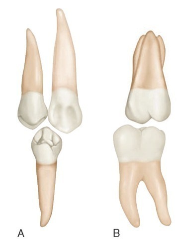
The crowns of the teeth are formed in such a way that cusps and ridges may slide over each other without mutual interference. In addition, the crowns of the teeth are turned on the root bases to accommodate the angled movement across their opponents (Figure 16-46).
Figure 16-45 Differences in jaw position between centric relation and the intercuspal position/centric occlusion position. A, Centric relation. B, Intercuspal position/centric occlusion.
The cusp tip of the mandibular canine moves through the linguoincisal embrasure of the maxillary lateral incisor and canine. The cusp tip is often in contact with one of the marginal ridges making up the lingual embrasure above it. Its mesial cusp ridge is usually out of contact during the lateral movement. Its distal ridge contacts the mesial cusp ridge of the maxillary canine.
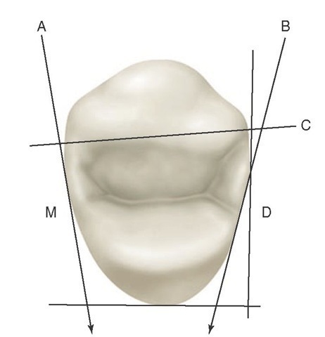
Figure 16-46 Occlusion of right maxillary first premolar with its distal (D) and lingual surfaces surveyed by a right angle distolingually (see Figure 9-15). A, Line following the angulation of the mesial surface (M), which is somewhat parallel with the vertical line of the right angle distally. This formation allows a proper contact relationship with the distal proximal surface of the maxillary canine; simultaneously, it cooperates with the canine in keeping the lingual embrasure design within normal limitations. B, Line demonstrating a more extreme angulation of the distal portion of the first premolar. This form allows cusp and ridge to bypass mandibular teeth over the distal marginal ridge surface of the maxillary tooth with normal jaw movements during lateral occlusal relations. C, Line aligned with mesiobuccal and distobuccal line angles, demonstrating the adaptation of the form of the buccal surface of the crown to the dental arch form without changing the functional position of the crown and root.
Figure 16-47 Lingual aspect. Interrelationship of cusp, ridges, and embrasures. A, Mandibular first premolar relation to maxillary canine and the first premolar approaching occlusal contact. B, Mandibular first molar relation to the maxillary first molar on the verge of occlusal contact.
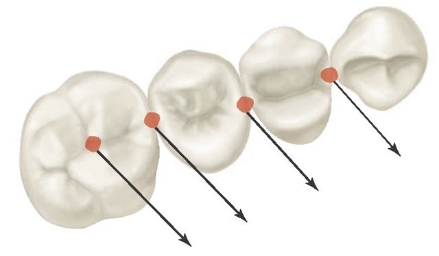
The cusp tip of the mandibular first premolar moves through the occlusal embrasure of the maxillary canine and first premolar (Figures 16-47 and 16-48). Its mesiobuccal ridge contacts the distal cusp ridge of the maxillary canine, and its distobuccal cusp ridge contacts the mesio-occlusal slope of the buccal cusp of the maxillary first premolar.
Figure 16-48 Arrows indicate path of left lateral movement of mandibular teeth over the maxillary teeth on nonworking side. Note the relationship of paths to morphological features of the teeth and embrasures.
The mandibular second premolar buccal cusp moves through the occlusal embrasure and then over the linguo-occlusal embrasure of the maxillary first and second premo-lars. Its mesiobuccal cusp ridge contacts the disto-occlusal slope of the buccal cusp of the maxillary first premolar, and its distobuccal cusp ridge contacts the mesio-occlusal slope of the buccal cusp of the upper second premolar.
The lingual cusps of all premolars are out of contact until centric relation is attained. Then the only lingual cusps in contact are those of the maxillary premolars, with the possible addition of the distolingual cusp of a mandibular second premolar of the three-cusp type. The molars have a more involved lateral occlusal relation because of their more complex design.
It has been noted previously, while describing the lateral occlusal relations of canines and premolars, that cusps, cusp ridges, sulci, and embrasures bear an interrelationship to each other. Cusps and elevations on the teeth of one arch pass between or over cusps and through embrasures or sulci. The tooth form and the alignment of the opposing teeth of both jaws make this possible. The cusps of the teeth of one jaw simply do not ride up and down the cusp slopes of the teeth in the opposing jaw. This explanation of the occlusal process has created wide misunderstanding. The cusp, ridge, fossa, and embrasure form of occlusion allow interdigitation without a "locked-in" effect. There is no clashing of cusp against cusp or any interference between parts of the occlu-sal surfaces if the development is proper.
