Chronologies of Human Dentition
The history of chronological studies demonstrates the difficulty in obtaining adequate documentation of the source of the data being presented. Many early tables and charts disagreed on the timing of chronological events. More precise information was needed to avoid injury to developing teeth during surgery on young children, especially related to the repair of cleft palate. One of the earliest of the widely used tables was that of Kronfeld.36 Kronfeld’s table was partly reprinted and altered by Schour and Massler37 and has a long history of subsequent development and compilations.10,19 Table 2-3 is an expanded and revised version that reflects the accumulated history of chronologies of tooth formation of Table 2-4. The latter table (Table 2-4) is the most commonly reprinted version of Kronfeld’s chronology.10
Even Table 2-3 and related chronologies36-40 have some deficiencies in sampling and collection methods, sufficient to prevent acceptance of chronologies that are not considered an ideal for standards of normal growth. The suggestions for revision made by Lunt and Law19 in their modification of the calcification and eruption schedules of the primary dentition (see Table 2-4) have been incorporated into the Logan and Kronfeld chronology shown in Table 2-3. The problems associated with revising a table completely or "plugging in" revised data is apparent when it becomes necessary to make critical choices from among available sources, as illustrated in Tables 2-5 and 2-6 by Smith.14
Types of Chronologies
Chronologies of dental development reflect the use of different statistical methods to produce three different types of tooth formation data: age of attainment chronologies based on tooth emergence, age prediction chronologies based on being in a stage, and maturity assessment scales used to assess whether a subject of known age is in front of or behind compared with a reference population.
Stages of Tooth Formation
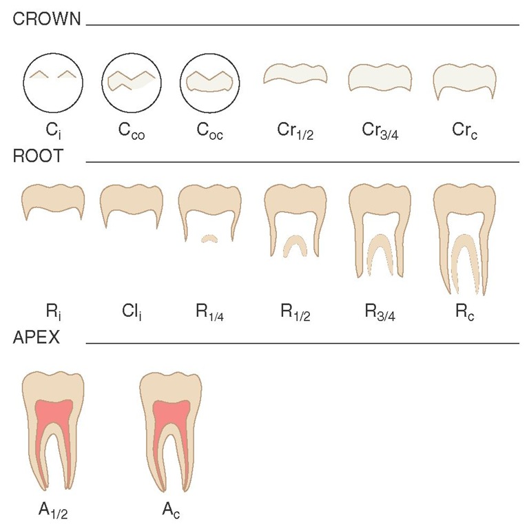
Radiographic studies of tooth formation have used at least three stages: beginning calcification, crown completion, and root completion. Nolla34 expanded the number of stages to 11 and Gleiser and Hunt to 13,44 which has served as the basis for several studies, including that of Moorrees et al,35 who defined 14 stages of permanent tooth formation (Figure 2-20). The 14 stages are not numbered but are designated by abbreviations (C = cusp; Cr = crown; R = root; Cl = cleft; A = apex) and subscripts (i = initiated; co = coalescence; oc = outline complete; and c = complete). Moorrees et al45 studied the development of mandibular canines and provided normative data.
Age of Attainment
The age of attainment of a growth stage is not easily determined, because in a proportion of the cases observed the attainment of the stage has not occurred, and in others the stage is over. Several procedures to answer the question of when a growth stage did occur have been used to construct chronologies of tooth formation, but many of these methods lead to chronologies that are not comparable for various reasons, including the problem of having fundamentally different underlying variables. Thus, the major statistical methods used to construct different statistically based chronologies of tooth formation relate to fundamentally different variables and should be used for different purposes.14 Such tables attempt to answer when the event usually happens—that is, at what age does the transition into the stage occur?14
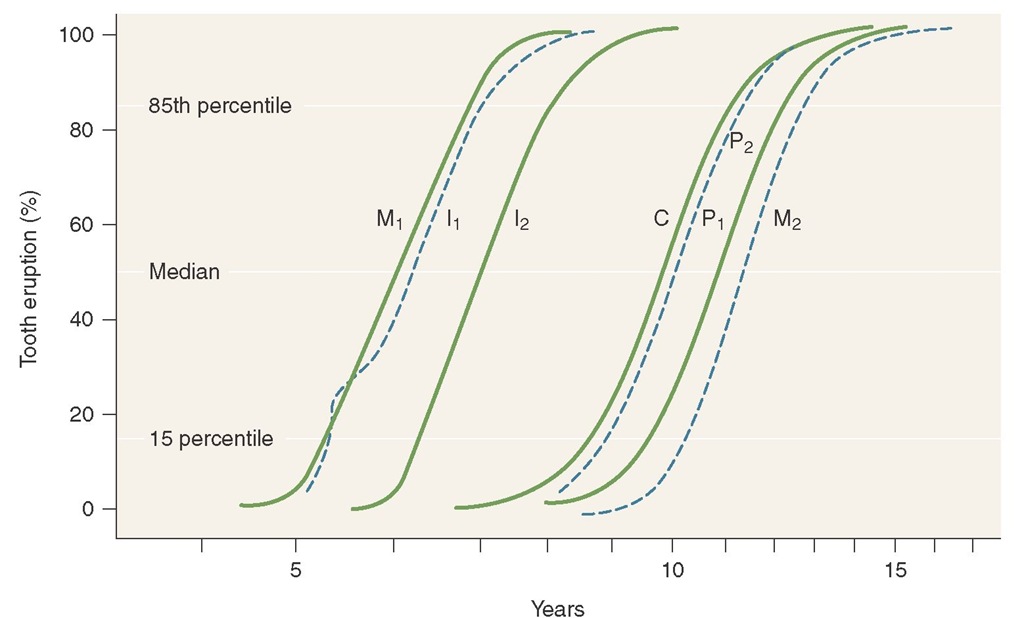
Age of attainment type of chronologies may be produced by cumulative distributive functions or probit analysis14 and by the average of age at first appearance less one half interval between examinations.46 Cumulative distributive functions, which have been used by a number of investigators (including Garn et al10,47 and Demirjian and Levesque48), appear to be the best method of determining the age of attainment.49 An example of this type of chronology is a schedule of tooth emergence as illustrated in Figure 2-21, where the proportion of subjects that have attained a particular stage is plotted against the midpoint of each age group. A chronology of age of attainment of tooth formation for females is shown as an example of this type of chronology (Table 2-7). Age of attainment schedules are useful clinically when it is necessary to avoid damage to developing teeth during treatment.
Table 2-3 Chronology of Human Dentition*
il, Central incisor; i2, lateral incisor; c, canine; ml, first molar; m2, second molar; 11, central incisor; 12, lateral incisor; C, canine; P1, first premolar; P2, second premolar; Ml, first molar; M2, second molar; M3, third molar.
*Part of the data from chronology of the growth of human teeth in Schour and Massler,37 modified from Kronfeld36 for permanent teeth and Kronfeld and Schour38 for the deciduous teeth. From Logan and Kronfeld,39 slightly modified by McCall and Schour (in Orban40) and reflecting other chronologies: a, Lysell et al13; b, Nomata41; c, Kraus and Jordan42; Lunt and Law19; d, mean age in months, ±1 standard deviation.
Table 2-4 Modified Table of Human Dentition*
SD, Standard deviation.
^Modification of the table from Chronology of the Human Dentition (Logan and Kronfeld,39 slightly modified by McCall and Schour [in Orban40]), suggested by Lunt and Law,19 for the Calcification and Eruption of the Primary Dentition.
!From Kraus and Jordan,42 pp. 107, 109, and 127 (except variation ranges of lateral incisors and canines). ^Modified from Lysell et al.13
Variation ranges of lateral incisors and canines from Nomata.41 (Fetal length-to-age conversions were made; no values are available for late onset in mandibular lateral incisors and canines, since all values from Nomata’s data are earlier than the mean values from Kraus and Jordan.42) Fetal length-to-age data from Patten.43
Age Prediction
Chronologies of tooth formation based on the average age of subjects in a stage of development have been suggested by several investigators.44,50-52 Although these kinds of chronologies are better suited for age prediction than age of attainment, no chronology is ideal for this purpose, and an alternate strategy suggested by Goldstein53 has been used by Smith14 to calculate age prediction tables for mandibular tooth formation. Values for predicting age in females are presented in Table 2-8. Such a table is useful to find out the age of the individual. In this schedule, each tooth is assessed independently, and the mean of all available ages is assigned as the dental age. In Table 2-8 the age related to a stage reflects the midpoint between mean age of attainment of the current stage and the next one. Age prediction chronologies are used for assessing unknown ages of patients and for forensic and archaeological applications.
Maturity Assessment
Chronologies of maturity assessment have been based on mean stage for age, where stages, rather than subject ages, are averaged.34,54 However, to avoid problems associated with the calculation of mean age or mean stage, maturity scales have been designed by several investigators, including Wolanski,55 Demirjian et al,29 Healy and Goldstein,56 and Nystrom et al.57 Such scales are useful when maturity is assessed for subjects of known age but are not designed for anthropological or forensic applications.14
Table 2-5 Available Values for Prenatal Formation of Primary Teeth
di1, Deciduous central incisor; di2, lateral incisor; dc, canine; dm1, first molar; dm2, second molar.
*Earliest age at which mineralization is seen through age at which 100% of the sample shows initial mineralization.
![]() These values are based on "tooth ring analysis"; they remain almost the only nonpictorial data available for deciduous incisors.14
These values are based on "tooth ring analysis"; they remain almost the only nonpictorial data available for deciduous incisors.14
Figure 2-20 Stages of permanent tooth formation. See text for definition of abbreviations.
Table 2-6 Ages for Postnatal Development of Mandibular Deciduous Teeth Expressed in Decimal Years
SD, Standard deviation; dil, central incisor; di2, lateral incisor; dc, canine; dml, first molar; dm2, second molar. *These data comprise an age-of-attainment schedule.
fThe basis of these values may be some combination of "tooth ring analysis" and observation of an infant sample; no other values could be located for deciduous incisors in studies with documented methods.14
Figure 2-21 Age of attainment of growth stage using a cumulative distribution function in which data represent tooth emergence.![]() First molar;
First molar;![]() central incisor;
central incisor;![]() lateral incisor;
lateral incisor;![]() canine;
canine;![]() first premolar;
first premolar;![]() second premolar;
second premolar;![]() second molar.
second molar.
Table 2-7 Mean Age of Attainment of Developmental Stages for Females (Permanent Mandibular Teeth)*
Il, Central incisor; I2, lateral incisor; C, canine; Pl, first premolar; P2,second premolar; Ml, first molar; M2, second molar; M3, third molar;![]() Cusp initiated;
Cusp initiated;![]() cusp coalesced;
cusp coalesced;![]() cusp outline completed;
cusp outline completed;![tmp182-194_thumb[2][2][2] tmp182-194_thumb[2][2][2]](http://what-when-how.com/wp-content/uploads/2012/05/tmp182194_thumb222_thumb.jpg) crown half completed;
crown half completed;![]() crown three quarterscompleted;
crown three quarterscompleted;![]() crown completed;
crown completed;![]() root initiated;
root initiated;![]() initial cleft formation;
initial cleft formation;
![]() root one fourth completed;root half completed;
root one fourth completed;root half completed;![tmp182-200_thumb[2][2][2] tmp182-200_thumb[2][2][2]](http://what-when-how.com/wp-content/uploads/2012/05/tmp182200_thumb222_thumb.jpg) root two thirds completed;root three fourths completed;
root two thirds completed;root three fourths completed;![]() root completed;apex half completed;
root completed;apex half completed;![]() apex completed.
apex completed.![tmp182-203_thumb[2][2][2] tmp182-203_thumb[2][2][2]](http://what-when-how.com/wp-content/uploads/2012/05/tmp182203_thumb222_thumb.jpg)
![tmp182-204_thumb[2][2][2] tmp182-204_thumb[2][2][2]](http://what-when-how.com/wp-content/uploads/2012/05/tmp182204_thumb222_thumb.jpg)
![tmp182-205_thumb[2][2][2] tmp182-205_thumb[2][2][2]](http://what-when-how.com/wp-content/uploads/2012/05/tmp182205_thumb222_thumb.jpg)
*Values interpolated from Moorrees et al35; all ages in years.14
Table 2-8 Values for Predicting Age from Stages of Permanent Mandibular Tooth Formation—Females*
See Table 2-7 for definitions of tooth designations and developmental stages. *Values interpolated from Moorrees et al35; all ages in years.14
Table 2-9 Primary Crown and Root Formation: Onset and Duration* Mandibular Deciduous Dentition
*Data partly derived from Smith.14 Smith’s sources included Moorrees et al,35,45 Sunderland et al,58 Anderson et al,46 Kronfeld,36 Lysell et al,13 and Hume.59
![]() are general deciduous tooth designations used by some anthropologists to indicate the central and lateral incisors, canine, first molar, and second molar, respectively.
are general deciduous tooth designations used by some anthropologists to indicate the central and lateral incisors, canine, first molar, and second molar, respectively.


