Arterial Supply to the Teeth
The arteries and nerve branches to the teeth are mere terminals of the central systems. This topic must confine itself to dental anatomy and the parts immediately associated, and references will therefore be made only to those terminals that supply the teeth and the supporting structures.
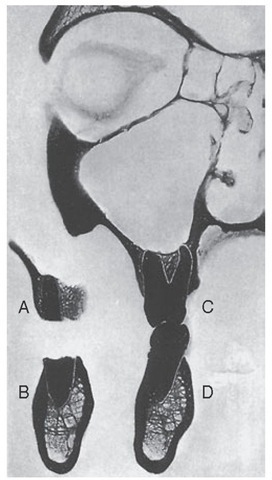
Figure 14-31 Second molar regions, showing relations of distobuccal root (A), distal root (B), and mesiobuccal (C) and lingual (D) roots with mesial half.
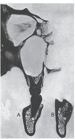
Figure 14-32 Third molar regions. A, Mesial root; B, apex of distal root; note deep groove for descending palatine artery.
INTERNAL MAXILLARY ARTERY
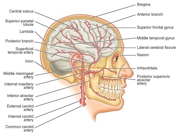
The arterial supply to the jaw bones and the teeth comes from the maxillary artery, which is a branch of the external carotid artery (see Figure 14-35). The branches of the maxillary artery that feed the teeth directly are the inferior alveolar artery and the superior alveolar arteries (Figure 14-35).
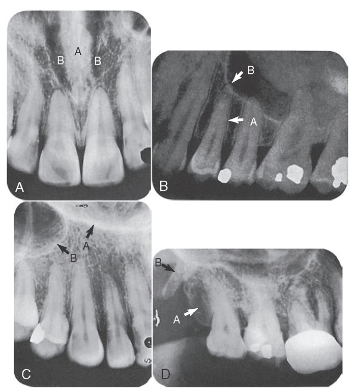
Figure 14-33 I A, Radiograph of central incisor region visualizing the nasal septum (A) and fossae (B). B, Radiograph demonstrating the normal appearance of the lamina dura (A) and the periodontal membrane (B). C, Radiograph depicting the Y (inverted) formed by the junction of the lateral wall (A) of the nasal fossa and the antemedial wall (B) of the maxillary sinus. D, Radiograph visualizing the tuberosity of the maxilla (A) and the hamular process of the sphenoid bone (B).
INFERIOR ALVEOLAR ARTERY
The inferior alveolar artery branches from the maxillary artery medial to the ramus of the mandible. Protected by the sphenomandibular ligament, it gives off the mylohyoid branch, which rests in the mylohyoid groove of the mandible and continues along on the medial side under the mylohyoid line. After giving off the mylohyoid branch, it immediately enters the mandibular foramen and continues downward and forward through the mandibular canal, giving off branches to the premolar and molar teeth. In the vicinity of the mental foramen, it divides into a mental and an incisive branch. The mental branch passes through the mental foramen to supply the tissues of the chin and to anastomose with the inferior labial and submental arteries. The incisive branch continues forward in the bone to supply the anterior teeth and bone and to anastomose with those of the opposite side.
The anastomosis of the mental and incisive branches furnishes a good collateral blood supply for the mandible and teeth.4
In their canals, the inferior alveolar and incisive arteries give off dental branches to the individual tooth roots for the supply of the pulp and periodontal membrane at the root apex (see Figure 5-12). Other branches enter the interdental septa, supply bone and adjacent periodontal membrane, and terminate in the gingiva. Numerous small anastomoses connect these vessels with those supplying the neighboring alveolar mucosa.
Figure 14-33 II A, Radiograph of the medial palatine suture, the appearance of which might be interpreted as a fracture. Radiographs visualize various extensions of the maxillary sinus. B, Alveolar extension. C, Tuberosity extension. D, Radiograph in which the canal for a superior alveolar artery is seen. E, Radiograph showing typical superimposition of the coronoid process: A, of the mandible on the tuberosity; B, of the maxilla.
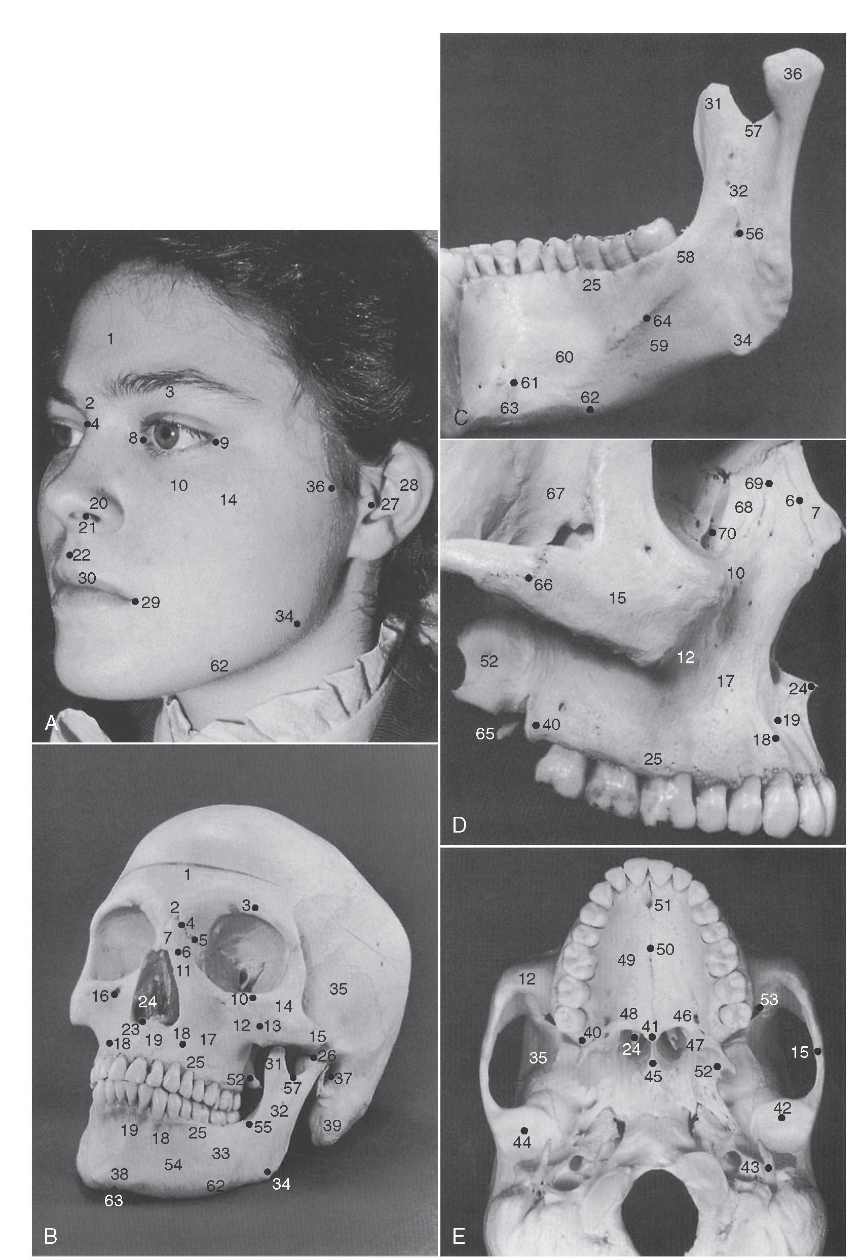
Figure 14-34 Surface landmarks. Various dental structures in the patient’s face can be quickly located by means of surface landmarks. Surface landmarks are identified in A. The photograph of the bony skull (B) was made from the same angle of view. Features of both are numbered and identified in the legend. The medial aspect of the mandible (C) shows anatomical details not clearly seen in the other illustrations. The maxilla and zygoma are shown in D. The bony anatomy of the hard palate and its adjoining structures is shown in E.
|
1, Frontal bone (forehead) |
25, |
Alveolar process |
49, |
Palatine process of maxilla |
|
2, Glabella |
26, |
Temporomandibular articulation |
50, |
Midpalatal suture |
|
3, Supraorbital ridge (superciliary ridge) |
27, |
Tragus of ear |
51, |
Incisive foramen |
|
4, Frontonasal suture (bridge of nose) |
28, |
Auricula |
52, |
Lateral pterygoid plate of sphenoid bone |
|
5, Maxillofrontal suture |
29, |
Labial commissure |
53, |
Inferior orbital fissure |
|
6, Maxillonasal suture |
30, |
Vermilion border of lip |
54, |
Mental foramen |
|
7, Nasal bone |
31, |
Coronoid process of mandible |
55, |
Oblique line |
|
8, Medial canthus |
32, |
Ramus of mandible |
56, |
Mandibular foramen |
|
9, Lateral canthus |
33, |
Body of mandible |
57, |
Mandibular notch |
|
10, Infraorbital ridge |
34, |
Gonial angle of mandible |
58, |
Internal oblique ridge |
|
11, Frontal process of maxilla |
35, |
Infratemporal fossa |
59, |
Submandibular fossa |
|
12, Zygomatic process of maxilla |
36, |
Condyle |
60, |
Sublingual fossa |
|
13, Zygomaticomaxillary suture |
37, |
External acoustic meatus |
61, |
Genial tubercle |
|
14, Zygomatic bone (cheekbone) |
38, |
Mental protuberance |
62, |
Inferior border of mandible |
|
15, Zygomatic arch |
39, |
Mastoid process of temporal bone |
63, |
Symphysis |
|
16, Infraorbital foramen |
40, |
Maxillary tuberosity |
64, |
Mylohyoid line |
|
17, Canine fossa |
41, |
Posterior nasal spine |
65, |
Hamular process of sphenoid bone |
|
18, Canine eminence |
42, |
Articular eminence |
66, |
Zygomaticotemporal suture |
|
19, Incisive fossa |
43, |
Styloid process of temporal bone |
67, |
Greater wing of sphenoid bone |
|
20, Nasal ala |
44, |
Mandibular fossa |
68, |
Lacrimal bone |
|
21, Nares |
45, |
Vomer |
69, |
Maxillolacrimal suture |
|
22, Philtrum |
46, |
Greater palatine foramen |
70, |
Lacrimal fossa |
|
23, Anterior nasal spine |
47, |
Lesser palatine foramen |
||
|
24, Inferior nasal concha |
48, |
Palatine bone |
Figure 14-35 Projection of maxillary artery and its branches in relation to brain, skull, and mandible, including the teeth.
