(Fig. 7.25). Starting in 1971, the military used a vaccine to
and evolutionary history of polyomaviruses and papillo-
prevent the disease. This vaccine consisted of live nonat-
maviruses are distinct and they are now classified into two
tenuated virus that was encapsulated in coated capsules and
distinct families.
taken orally. Infection of the enteric tract is asymptomatic
To date, 14 different polyomaviruses have been described
for these viruses under these conditions and results in a
and others probably exist. These viruses infect warm-blooded
good antibody response, presumably including mucosal
vertebrates. Mammalian viruses include viruses infecting
antibodies to protect the respiratory tract. Use of this vac-
humans, monkeys, cattle, rabbits, rats, mice, hamsters, while
cine reduced adenovirus-specific disease rates in trainees
bird viruses infecting parakeets, geese, and other birds are
by 95 to 99% and reduced the total respiratory disease rates
known. The majority of infections of their native host by the
by 50 to 60%, illustrating the importance of Ad4 and Ad7
mammalian viruses are asymptomatic and the viruses estab-
as causative agents of severe respiratory disease. The vac-
lish a lifelong infection. Many can cause tumors in nonnative
cine is no longer available, however. It was made by a sin-
hosts, however, and may be associated with tumors in their
gle manufacturer whose facilities for making the vaccine
native hosts, and have been intensively studied as models for
became outdated and needed upgrading. The army was
tumor induction. In contrast, the bird viruses cause severe
unwilling or unable to find a mechanism to help with the
disease with a high mortality rate in their native host. A par-
cost of the upgrade, and since the military was the sole pur-
tial listing of the members of the family Polyomaviridae is
chaser of the vaccine, production was discontinued in 1995.
given in Table 7.12. The two best studied viruses are mouse
The last of the vaccine was exhausted in 1999 and epidem-
polyomavirus (MPyV), so called because it causes many
ics of severe adenovirus respiratory disease in recruits are
(poly) different kinds of tumors (omas) in mice, and sim-
once again a serious problem.
ian virus 40 (SV40), which infects monkeys. These viruses
Adenoviral epidemics such as have occurred in mili-
share about 60% nucleic acid sequence identity. SV40 was
tary recruits are rare in the civilian sector but do occur. As
first recognized as a contaminant in monkey kidney cultures
one example, there was an epidemic of Ad11 in 1997 in a
used for the production of poliovirus vaccine. Two polyoma-
South Dakota job-training facility. Like a military camp,
viruses for which humans are the natural host are known, BK
this facility had young adults housed in crowded condi-
virus and JC virus. SV40 also circulates in humans, possibly
tions with regular multiple introductions of new suscep-
as a result of its introduction with the contaminated polio-
tible persons. Illness with severe morbidity also occurs
virus vaccine. These three viruses have received increasing
in pediatric populations, school environments, and health
attention as disease agents.
care settings.
The structures of the two polyomaviruses SV40 and
Adenoviruses are being developed as vectors to immu-
MPyV have been solved to atomic resolution (Fig. 2.10).
nize people against other viruses and for gene therapy. Two
They are icosahedral viruses 45 nm in diameter with pseudo-
approaches are being used. One is to use the military experi-
T=7 symmetry (Figs. 2.1 and 2.5). The capsid consists of
ence with the Ad4 and Ad7 oral vaccines to develop vaccines
72 pentamers of structural protein L1, and each pentamer
that could be delivered safely using this route. A second
contains one molecule of either structural protein L2 or L3.
approach is to disable the virus by deleting some of the early
The polyomavirus genome is a circular dsDNA molecule
genes. Such an attenuated virus has been used in clinical tri-
that is 5 kb in size. In the virus it is complexed with cellular
als for treatment of cystic fibrosis. In these trials, virus that
histones to form a supercoiled minichromosome.
expresses the gene that is defective in such patients is deliv-
ered directly to the lungs. Trials that use adenoviruses in an
The Early Genes
attempt to treat other diseases are also ongoing. The use of
adenovirus for gene therapy has not been very successful to
Transcription maps of SV40 and MPyV are shown in
date, and the death of a patient in a gene therapy trial from
Fig. 7.26, in which the circular genomes are represented as
adenovirus led to retrenchment in this area. This topic is dis-
linear structures for ease of presentation. The genomes are
cussed in more detail in Chapter 11.
divided into two domains, an early domain that is transcribed
from one DNA strand and a late domain transcribed from
the other strand. An origin (Ori) is present at the junction
FAMILY POLYOMAVIRIDAE
of the two domains. Ori serves as an origin of replication for
DNA replication and a transcription start site for transcription
In the past the polyomaviruses and the papillomaviruses
by RNA polymerase II. It also contains enhancer ele-
were considered to be two subfamilies within the family
ments.
Papovaviridae. The name papova came from papilloma
Following infection, the viral DNA is transported to the
virus/polyoma virus/simian vacuolating virus (= SV40),
nucleus, whether as part of a virion or subviral particle or
three characteristic members of the enlarged family.
as free DNA is not clear, and early mRNA is transcribed by
However, the mode of replication, genome organization,
cellular RNA polymerase. The MPyV or SV40 promoters
TABLE 7.12 Polyomaviridae a
Virus name
Usual
Genus/members
abbreviation
host(s)
Transmission
Disease
Polyomavirus
PMLb-like disease in rhesus monkeys and
Simian virus 40
SV-40
Monkeys
immunocompromised macaques; may cause
Reactivation of persistent
childhood brain tumors in humans
infection, virus shedding
in urine, contact, aerosols,
Murine polyoma
MPyV
Mice
Tumors when inoculated into newborn mice, virus
sexual transmission
persists in the kidney
Bovine polyomavirus
BPyV
Cattle
Common infection, persists in the kidney
BK polyomavirus
BKPyV
Humans
Transmission as above plus
Common infection of early childhood; virus persists;
tissue transplantation in humans
tumors in immuncompromised humans
Common infection of late childhood; causes PMLb in
JC polyomavirus
JCPyV
Humans
immunocompromised hosts
Budgerigar fledgling
BFPyV
Parakeets
Fatal illness in fledgling parakeets
polyomavirus
Plus other viruses infecting monkeys, hamsters, and rabbits
a
Most polyomaviruses are worldwide in distribution.
b
PML, progressive multifocal leukoencephalopathy.
are strong promoters, active in many cells. Because of this,
Binding of large T antigen to the Ori regulates its own
the SV40 promoter has been used to drive expression of
production. Binding is also required for DNA replication.
foreign genes in many different expression systems. Early
Binding of SV40 large T antigen to Rb and p53, as well
mRNA transcribed from the viral genome terminates about
as to p107 and p300, is illustrated in Fig. 7.22. Binding to these
halfway around the genome and is differentially spliced to
tumor-suppressor proteins results in continued cell cycling
yield several mRNAs (Fig. 7.26). The translation products of
and transformation of the cell. Binding to Rb and its rela-
these mRNAs are called T antigens (tumor antigens) because
tives p107 and p300 releases transcription factors required
their expression in the absence of productive viral infection
for cell cycling, of which the best studied and perhaps most
leads to cell transformation and the formation of tumors in
important are members of the E2F family. p53 is a transcrip-
animals. The two major SV40 proteins are called the small
tion factor whose activity can prevent DNA synthesis, cause
t antigen and the large T antigen, whereas the three major
G2 and G1 growth arrest, and induce apoptosis. As described
MPyV antigens are called small t, middle T, and large T anti-
earlier, cell cycling induces a state suitable for viral DNA
gens. In addition, a 17k T antigen has been reported in SV40
replication. Expression of large T in the absence of other
and a short-lived tiny t antigen has been reported for MPyV.
SV40 proteins is sufficient to transform a cell, although such
The large T antigens (size: about 700 amino acids) are
a transformed cell does not express the full complement of
multifunctional proteins that interact with viral promoters
transformed attributes unless small t is also expressed.
and several cellular proteins. A schematic representation of
The activities in MPyV virus T antigens are distributed
the large T antigen of SV40 that illustrates the locations of
somewhat differently. Polyoma large T antigen binds Rb
many of its activities within the protein sequence is shown
but does not bind p53. Binding to Rb induces cellular DNA
in Fig. 7.27. These activities are differentially regulated by
synthesis. Cell transformation is a function of middle T anti-
phosphorylation of serine and threonine residues, as shown.
gen. Middle T antigen binds to src, a known proto-oncogene
The large T antigen possesses a nuclear localization signal
located at the cell surface, and activates its kinase activity
that causes it to be transported to the nucleus, a domain that
(Fig. 7.28). The phosphorylated middle T antigen interacts
binds to sequence elements within Ori, phosphorylation
with other cellular factors and induces cell transformation,
sites in two widely separated regions that serve to regulate
and it has been shown that expression of middle T antigen
its activities, domains that interact with DNA polymerase α-
alone is sufficient to transform a cell.
primase, a domain that controls the host range of the virus, a
The oncoproteins of these small DNA viruses are excel-
domain that has DNA helicase activity, and domains that bind
lent examples of how a viral protein may be multifunctional
cellular proteins that play regulatory roles in cell cycling,
and mediate a variety of functions important for the outcome
among others.
of infection.
A. SV40 (5243 bp)
Ori
Ori
VP3
Ag
VP2
An
Late Transcripts
Ag
An
VP1
PL
DNA
PR
NTR
An
Small t
Regulatory
Early Transcripts
Region
An
Large T-antigen
Scale (bp)
0
1000
2000
3000
4000
5000
B. Mouse Polyoma (5297 bp)
Ori
Ori
Large T-antigen
An
An
Middle
T antigen
Early Transcripts
Enhancer
An
Small t
NTR
P
R
DNA
P
L
An
VP1
An
Late Transcripts
VP2
An
VP3
0 Scale (bp)
5000
400
3000
2000
1000
0
P Early gene promoter
Ori
ORF 1
ORF 4
Initation codon
Origin of replication
R
P Late gene promoter
Termination codon
ORF 2
ORF 5
NTR (regulatory
L
region or enhancer)
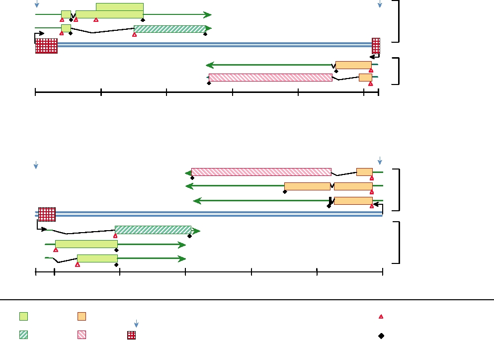
FIGURE 7.26 Comparisons of the genome organizations of two polyomaviruses: SV40 and mouse polyoma. Both are
circular genomes which have been linearized at the origin of replication (Ori) for ease of presentation. The genome of
mouse polyoma virus is shown here in the opposite sense (right to left) to that of SV40. Ag is the agnoprotein. Redrawn
after Brady and Salzman (1986).
DNA Replication
The Late Genes
Large T antigen binds to specific sites within the Ori
Large T antigen also regulates the transcription of late
to promote replication of the viral genome. Binding first
mRNAs, which are transcribed from the opposite strand as the
unwinds the DNA. Then T antigen associates with repli-
early mRNAs (Fig. 7.26). Differential splicing leads to two
cation protein A followed by DNA polymerase α-primase
mRNAs in SV40 and three mRNAs in polyoma. One mRNA
to form an initiation complex. Association with primase is
is translated into VP1, the major virion structural protein. In
species specific. As a result, SV40 productively infects only
SV40 the second mRNA is translated into both VP2 and VP3,
monkey cells and mouse polyomavirus infects only mouse
whereas in polyoma virus VP2 and VP3 are translated from
cells. After initiation by primase, DNA polymerase takes
different mRNAs. VP3 is a truncated form of VP2, consisting
over and replication proceeds. DNA synthesis is bidirec-
of the C-terminal 60% or so of VP2. Both are minor com-
tional and when the replication forks meet about halfway
ponents of the virion. VP2 is myristylated and may serve an
round the molecule, the daughter genomes separate, aided
entry function. The three structural proteins are transported to
by topoisomerase II (Fig. 1.9A).
the nucleus and assembly of virions takes place there.
Large T Protein
SST
SS
NH
COOH
120 124
677
679
123
0
100
200
300
400
500
600
700
Amino acids
N
ATP
Polα
Zn
L
Ori binding
binding
HR
S
Functional
ATPase
domains of
p53 binding, Polα binding
large T
Rb
X
DNA helicase activity
Phosphorylation
by unknown kinases
ATP
N
Polα
binding
Ori binding
Zn
L
HR
S
Activities of
ATPase
phosphorylated
p53 binding, Polα binding
large T
X
Rb
DNA helicase activity
Dephosphorylation by PP2A
SS
SS
ATP
N
Polα
binding
Ori binding
Zn
L
HR
S
Activities of
ATPase
dephosphorylated
p53 binding, Polα binding
large T
X
Rb
DNA helicase activity
Initiation of viral DNA Synthesis
N
Activity enhanced
Host range helper function
Nuclear localization signal
L
HR
S
Activity reduced
Zn finger
Zn
Phosphate group
domain
Activity unchanged
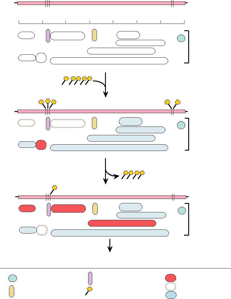
FIGURE 7.27 Functional domains of SV40 large T antigen. The top panel illustrates the location of various functional
domains and of the serine and threonine residues that are phosphorylated. The second panel shows the functions of fully
phosphorylated large T antigen. The bottom panel shows the activities of large T singly phosphorylated on threonine 124,
which can bind to the origin of replication to initiate DNA synthesis. In the middle and lower panels, blue domains are
unchanged in activity, gray domains are reduced in activity, and red functions are strongly increased. Adapted from Berg
and Singer (1997), Figures 1.23 and 1.24 and Fields et al. (1996), Figure 6 on p. 2011.
Cell Membrane
P
P S
c-src protein
P S
Y
SH2
Middle T antigen
P13K protein
Association of middle T with c-src and
activation of c-src tyrosine kinase
Cell Membrane
P
S
P
P S
Y
AT P
Middle T antigen
SH2
P13K protein
Phosphorylation of middle T by c-src on Y
Cell Membrane
P
P S
P S
Y
P
SH2
Middle T antigen
P13K protein
Transformation
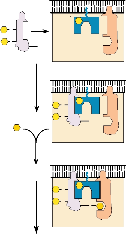
FIGURE 7.28 Interactions of polyoma middle T antigen with membrane proteins to initiate cellular transformation.
Phosphatidylinositol-3-kinase (Pl3K) and c-src are localized in the plasma membrane. The src kinase phosphorylates
middle T on a tyrosine residue (Y). This generates binding sites for a variety of other cellular signaling proteins such as
P13K. The resultant complex closely resembles an activated growth factor receptor bound to signal transduction proteins.
Serine residues (S) on middle T that are phosphorylated by other protein kinases are also indicated. Drawn from data in
DiMaio et al. (1998).
VP1, when expressed alone, assembles into virus-like
Polyomavirus Infection of Humans
particles. If VP2 and VP3 are coexpressed with VP1, they
The two well-known human polyomaviruses are BK
assemble into virus-like particles whose composition is the
virus and JC virus. These viruses were first isolated in
same as that of the virion.
1971, JC from the brain of a patient with progressive
Release of virions is an active process. Membrane vesicles
multifocal leukoencephalopathy (PML) and BK from the
form and transport virions to the cell surface, where they are
urine of an immunosuppressed renal transplant patient.
released. Infection by polyomaviruses is lytic. Expression of
They were named after the initials of the patients from
the late genes and assembly of progeny virions results in the
whom they were isolated. These two viruses share 75%
death of the cell.
TABLE 7.13 Virus-Coded Proteins of Primate Polyomaviruses
Number of amino acids
% Amino acid identity
Protein
JC
BK
SV40
JC/BK
JC/SV40
BK/SV40
Function
Late proteins
VP1
356
362
364
77.9
76.4
82.4
Major capsid protein, attaches to cellular receptors,
hemagglutination, HI and NT epitopes
VP2
344
351
352
78.8
73.4
78.5
Minor capsid protein
VP3
225
232
234
74.5
67.2
73.6
Minor capsid protein
Agnoprotein
71
66
62
59.1
45.0
53.2
Facilitates capsid assembly
Early proteins
Large T
688
695
708
86.6
72.0
73.9
Initiation of replication, stimulates host DNA
synthesis; modulates transcription,
transformation
Small T
172
172
174
79.6
67.8
69.5
Necessary for efficient viral DNA replication
Source: Adapted from Fields et al. (1996), p. 2030 and additional data from Walker and Frisque (1986).
nucleotide sequence identity. A comparison of the proteins
kidneys, in B lymphocytes, and, for JC virus, perhaps in the
encoded by JC, BK, and SV40 viruses is given in Table
brain. Reactivation may be brought about by immunosup-
7.13. Shown are the number of amino acids in each protein
pression or by factors such as pregnancy or diabetes, and
and the amino acid identity between any two of these
results in the excretion of virus in the urine. Reactivation
viruses. This table makes obvious the close relationship
of JC virus resulting from immune suppression is serious
among these viruses.
because it can replicate in oligodendrocytes, which produce
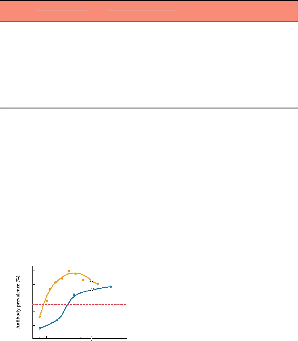
Most humans in the United States are infected with BK
myelin in the brain, and the death of these cells results in
virus before the age of 10 (Fig. 7.29). Infection with JC
PML. PML is a rare, subacute, demyelinating disease of the
virus usually occurs somewhat later, but by the age of 14 the
central nervous system that has a worldwide distribution. It
majority of the population has been infected. Primary infec-
is an infrequent complication of a wide variety of conditions,
tion with BK virus has been associated with mild respiratory
including Hodgkin's disease, chronic diseases such as tuber-
disease or cystitis (bladder infection) in young children, but
culosis, primary acquired immunodeficiency diseases such
most infections with either BK or JC virus are not associ-
as AIDS, or immunosuppression following organ transplant.
ated with illness. The viruses establish a latent infection that
The frequency of PML, once considered a rare disease, has
persists indefinitely, and the virus may reactivate after many
increased with the AIDS epidemic and PML is now recog-
years. The latent infection appears to be established in the
nized as one of the AIDS-defining illnesses--it occurs in
about 5% of all AIDS patients. PML may be more likely to
occur when immunosuppression is due to infection by HIV
because HIV-1 transactivates the JC late promoter. The dis-
100
BK virus
ease progresses rapidly and can lead to mental deterioration
and death within 36 months after onset.
80
Reactivation of BK virus can cause kidney disease. Such
reactivation as a result of immune suppression accompany-
JC virus
60
ing kidney transplantation is a leading cause of allograft
failure.
40
Polyomaviruses cause tumors in laboratory animals and
many attempts to associate BK or JC virus with human
20
cancer have been made. Association of these viruses with
many human malignancies has been reported, but proof that
20
4
8
12
16
>50
they cause these malignancies is still lacking. Both viruses
persistently infect most humans, and the presence of one
Age in years
of these viruses in association with a tumor cell does not
FIGURE 7.29 Prevalence of antibodies to BK and JC viruses in humans
prove causality, as the tumor cell could provide a suitable
in the United States as a function of age. From Fields et al. (1996), p. 2039.
Search WWH :