FAMILY ADENOVIRIDAE
and HHV-8. Intriguingly, Kaposi's sarcoma is 15-fold more
common in homosexual male AIDS patients than in patients
who acquired HIV by a nonsexual route. HHV-8 is also asso-
Adenoviruses are widespread viruses of mammals and
ciated with body cavitybased lymphoma, a lymphoid tumor
birds, but a few of the known viruses are able to infect rep-
in some AIDS patients, and with multifocal Castleman's dis-
tiles or frogs. The virions are a T=25 icosahedron, 7090 nm
ease, a rare lymphoproliferative disorder. In common with a
in diameter, with fibers 9 to 77 nm in length projecting from
number of other herpesviruses, HHV-8 encodes factors that
the 12 fivefold axes of the icosahedron (Figs. 2.1 and 2.12).
stimulate cells to divide, that interfere with the host immune
Virions contain about a dozen proteins, of which 4 are present
response, and that block apoptosis. It also produces a protein
in the core. The major structural proteins are a hexon protein
in latently infected cells that tethers the viral DNA to mitotic
called II, three copies of which form a hexon, of which there
chromosomes.
are 240 in the virion, and a penton protein called III, five
copies of which form a penton base, of which there are 12 in
the virion. The genome of adenoviruses is a linear dsDNA of
Monkey B Virus
size 26 to 45 kb. A terminal protein that served as a primer
Many herpesviruses infect vertebrates other than humans,
during DNA replication is covalently attached to the 5¢ end
but only one nonhuman herpesvirus is known to be highly
of both strands.
pathogenic for humans. Cercopithecine herpesvirus 1 or
Adenoviruses are named after adenoids, a glandlike col-
monkey virus B is indigenous to Old World monkeys in the
lection of lymphoid tissue in the nasopharynx. Many human
genus Macaca. In its native host it causes a disease that is
adenoviruses establish a long-term infection in this tissue and
similar to that caused by HSV-1 in humans. A latent, lifelong
adenoviruses were first isolated from human adenoids. Four
infection is established in the monkey that seldom leads to
genera are currently recognized. The genus Mastadenovirus
serious illness. Sporadic reactivation of the virus occurs that
contains viruses that infect only mammals and the genus
results in the formation of vesicular lesions, particularly on
Aviadenovirus contains viruses that infect only birds, whereas
the tongue and cheeks. However, infection of humans or of
the genera Atadenovirus and Siadenovirus contain viruses that
a number of monkeys other than macaques results in a very
infect a wide variety of vertebrates (Table 7.10). Most viruses
serious neurological disease that has a high fatality rate. As
are species specific and in general will only undergo a com-
described earlier, HSV-1 is also neurotropic and occasion-
plete replication cycle in cells isolated from their native host.
ally causes fatal encephalitis.
Fifty-one human adenoviruses have been distinguished
B virus has usually been transmitted to humans through
on the basis of serological reactivity--an adenovirus is con-
the bite of a monkey in which infectious virus was present
sidered distinct if it resists neutralization by antisera against
in the saliva. However, transmission has also occurred by
the other known adenoviruses. All belong to the genus
other means. In at least one case transmission resulted from
Mastadenovirus and have a genome size of 3036 kb. The
contact of infectious material with the eye and two cases are
51 viruses are simply numbered in order of their isolation
thought to have resulted from exposure to aerosols. One case
and are often referred to as Ad1, Ad2, etc., or more formally
of transmission of virus from an animal worker to his wife
as HAdV-1, etc., to distinguish them from adenoviruses
has also been documented. The majority of human infec-
that infect other species. The human viruses were originally
tions have resulted in fatal neurological disease, but some
divided into six subgroups on the basis of serological cross-
infections resulted in only mild disease and the establish-
reactions in a hemagglutination-inhibition assay. In this
ment of a latent infection. Acyclovir has been used to treat
assay, the ability of an antiserum to bind to the virus and
persons infected by B virus with apparent success, but the
prevent it from agglutinating red blood cells is examined.
number of cases is small and no controlled trials of efficacy
An antiserum against one of the viruses of subgroup A,
have been conducted.
for example, inhibits hemagglutination by all members of
Recurrence of herpes infections is often associated with
that subgroup but not by members of other subgroups. This
stress. Newly captured or shipped animals are subject to a
grouping correlated with a number of other properties of
great deal of stress, so that active infection in these animals
the viruses as well, such as their ability to form tumors in
is not uncommon. Animal handlers or researchers using
rodents. These original subgroups are now considered to be
these animals are at risk for the disease and must take proper
different adenovirus species, human adenovirus A through
precautions when handling macaques. Because of the dan-
F, with grouping relying on sequence identities where pos-
gers from B virus infection, most laboratories in the United
sible. Two viruses are considered to belong to the same spe-
States use only monkeys that lack antibodies to B virus, an
cies if they differ by less than 10% in their sequence, and
indication that they are not infected. Although this greatly
by this criterion the human adenovirus species may contain
reduces the risk of handling the animals, it does not elimi-
adenoviruses of other animals.
nate all risk because occasionally monkeys that are infected
Because they replicate to high titer in cultured human cells,
do not have detectable antibodies.
several human adenoviruses have been intensively studied,
TABLE 7.10 Adenoviridae
Genus/members
Serotypes
Usual host(s)
Disease in natural host
Mastadenovirus
Human adenovirus A
Types 12,18,31
Humans
Enteritis
Human adenovirus B
Types 3,7,11,14,16,21,34,35
Humans
Enteritis; military recruits' disease (3, 7, 14, 21); type 35
causes pneumonia in elderly and immunocompromised
humans
Human adenovirus C
Types 1,2,5,6
Humans
Respiratory infection in children
Human adenovirus D
Types 8,9,10,13,15,17, 19,20,
Humans
Enteritis
22-30,32,33,36-39,42-47
Human adenovirus E
Human type 4, simian types
Humans
Enteritis, pneumonia and upper respiratory disease in
22,23,24,25
military recruits
Human adenovirus F
Humantypes 40,41, simian type 19
Humans
Infant diarrhea
Murine adenovirus A
Mice
Bovine adenovirus A, B, C
Cattle
Asymptomatic or mild respiratory disease
Ovine adenovirus A, B
Sheep
Porcine adenovirus A, B, C
Swine
Canine adenovirus
Types 1 and 2
Dogs
Hepatitis (type 1)
Respiratory disease (type 2)
Aviadenovirus
Fowl adenovirus A, B, C,
Chickens, ducks
Hepatitis, bronchitis, duck hepatitis (rare)
D, E.
Goose adenovirus
Geese
Atadenovirus
Ovine adenovirus D
Sheep
Basis of some vectors for human gene therapy
Bovine adenovirus D
Cattle
Duck adenovirus A
Ducks
Egg drop syndrome
Siadenovirus
Frog adenovirus
Amphibians
Nonpathogenic
Turkey adenovirus 3
Turkey, pheasants,
Hemorrhagic enteritis in turkeys, marble spleen
chickens
disease in pheasants, splenomegaly in chickens
a
In general adenoviruses are transmitted by both aerosols and fomites and by the oralfecal route.
in particular Ad2, Ad5, and Ad12. Further interest has been
produce a set of early RNAs. Later, a set of late RNAs is
generated by the fact that, although they will not undergo a
produced. A transcription map of Ad2 is shown in Fig. 7.20.
complete replication cycle in rodent cells, they will infect
Transcription of early RNAs occurs from five promoters,
and transform rodent cells in culture. Members of HAdV-A
three on the so-called R strand and two from the L strand.
will also cause tumors in rodents at a high rate and members
The R strand is transcribed rightward on the chromosome
of HAdV-B at a moderate rate. Other human adenoviruses
as conventionally drawn, and the L strand is transcribed
cause tumors in rodents at a low or undetectable rate. There
leftward. There are two other promoters for transcription of
is no evidence that adenoviruses are associated with tumors
delayed early mRNAs. Multiple splicing of these transcripts
in humans, however. Because of the extensive background of
leads to the production of about 30 mRNAs. The proteins
information about them and the fact that they usually cause
translated from these early mRNAs are required for repli-
only minor illness, attempts are being made to use them as
cation of the viral genome. The E1A and E1B gene prod-
expression vectors for gene therapy (Chapter 11).
ucts are oncogenes that stimulate the cell to enter S phase
and thereby induce an ideal environment for the replication
of the viral DNA. Their mode of action is described later.
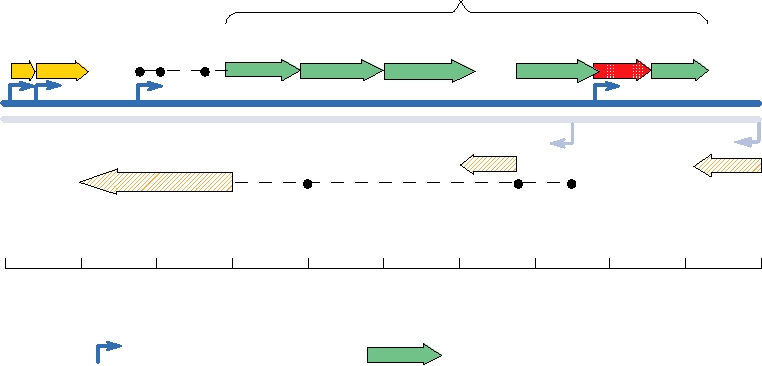
Transcription of Adenovirus mRNAs
Proteins from the E2 region are directly involved in repli-
Adenovirus replication takes place in the nucleus. After
cating adenovirus DNA and include a DNA polymerase, an
the entry of the infecting genome into the nucleus, the infect-
ssDNA-binding protein, and a precursor to the terminal pro-
ing genome is transcribed by host RNA polymerase II to
tein, which is involved in initiation of DNA replication. E3
irion structural proteins (green units)
E1A E1B
12
3
L1
L2
L3
L4
L5
E3
3
L
5
R 3
DNA
F
V
5
E4
E2B
E2A
Polymerase
72K - ssDNA binding
Map
Units
100
0
10
20
30
40
50
60
70
80
90
Promoter
Transcription Unit
IGURE 7.20
Genome organization of human adenovirus type 2. The double blue lines represent the two linear DNA
strands that make up the genome of 36 kbp. The genes have been mapped by superimposing an arbitrary scale of 100
map units. Each arrow represents a transcription unit composed of a nested set of spliced messages, transcribed in the
direction of the arrow. Black dots indicate short spliced sequences that form leaders. General functions of the various
transcription units are shown. Proteins in the E3 cluster interact with the host immune system, and E4 genes are involved
in DNA replication. The major late transcription unit includes the leaders "1," "2," and "3" and the L1, L2, L3, L4, and
L5 families of genes. Adapted from Wold and Golding (1991).
proteins modulate the host response to adenovirus infection,
Replication of the Viral DNA
and this region is nonessential in cultured cells. The func-
tions of the E3 proteins are described in Chapter 10. Region
Inverted terminal repeats that contain the origins
E4 encodes proteins involved in transcription and transport
of replication are present at the ends of the adenovirus
of viral mRNAs and in DNA replication.
genome. DNA synthesis is initiated at one of the two ends
Late genes are all transcribed beginning from a single pro-
and proceeds to the other end. There is no lagging strand
moter on the R strand, and transcription leads to an RNA prod-
synthesis and the partner of the strand being copied is
uct that is about 80% the length of the adenovirus genome.
displaced as an ssDNA (illustrated schematically in Fig.
Multiple splicing occurs to produce at least 18 different mRNAs
1.9C). The precursor of the terminal protein serves as a
that fall into five families, based on the use of five different
primer during initiation. It forms a complex in solution
polyadenylation sites. Each late mRNA has the same tripartite
with the adenovirus DNA polymerase, and it is assumed
leader, formed by splicing. The late mRNAs are translated into
that these two proteins bind to the origin of replication
the proteins required for the assembly of progeny virions.
as a complex during initiation of DNA replication. There
The multiple splicing events that occur during process-
are also binding sites within the terminal repeats for sev-
ing of adenoviral mRNA, especially the late mRNA, which
eral cellular proteins that stimulate the initiation of DNA
is made in abundance, led to the discovery of RNA splic-
synthesis. The first step in initiation is the covalent link-
ing by Phillip Sharp. Upon examination of adenoviral
age of dCMP, the first nucleotide in adenovirus DNA,
mRNADNA hybrids with an electron microscope, he
to the preterminal protein. Subsequent chain elongation
observed that regions of the genome were missing from the
requires the activity of the adenovirus DNA polymerase
mRNA transcripts. For his discovery of RNA splicing, he
and of the ssDNA binding protein, as well as of a cellular
was awarded a Nobel Prize, along with Richard Roberts, in
topoisomerase. The use of a protein primer eliminates the
1993 (Table 1.1).
need for a primase to initiate DNA synthesis with an RNA
In addition to the many genes transcribed by RNA
primer, and thus solves the problem of how to maintain
polymerase II, one or two adenovirus genes, called VA, are
the ends of the linear adenoviral DNA molecule during
transcribed by host RNA polymerase III. Short VA RNA
replication.
molecules are produced that are not translated. They func-
The products of the first round of replication are a dou-
tion to inhibit the host interferon system and the host RNAi
ble-strand progeny genome and a single-strand copy of one
system and will be described in Chapter 10, after the descrip-
of the two strands of the genome. Initiation of DNA syn-
tion of these host systems.
thesis can also occur on this ssDNA. It is proposed that a
panhandle structure is formed by the terminal repeats so that
from an mRNA that sediments at 12S) and 13S E1A (because
an origin of replication is present that is identical to that in
its mRNA sediments at 13S). Rb or retinoblastoma suscep-
the dsDNA. Copying this ssDNA renders it double stranded
tibility protein is a tumor suppressor. It was first identified
and completes the production of two copies of the dsDNA
because it is absent in patients suffering from retinoblastoma.
genome from the parental genome.
In its hypophosphorylated form, Rb binds a cellular transcrip-
tion factor called E2F and causes the cell cycle to arrest in G1.
Hyperphosphorylation of Rb causes it to dissociate from E2F.
Assembly and Release of Progeny Virions
Free E2F activates the transcription of genes that cause the
Progeny viruses are assembled in the nucleus from pre-
cell to enter S phase. The binding of Rb by E1A prevents it
assembled hexons and pentons (see Fig. 2.12). Viral DNA
from complexing with E2F, and E2F is thus free to induce the
is required for assembly. A packaging signal of about 260
cell to enter S phase. p107 and p130 are other members of the
nucleotides at the left end of the viral DNA leads to polar-
Rb family that also interact with E2F, as well as with cyclins
ized encapsidation starting from this end. During assembly,
and cyclin-dependent kinases, whose activities are disrupted
the viral protease cleaves at least four viral products, and
by binding to E1A. Binding of E1A to p300 appears to be
these cleavages are required to stabilize the particle and
an independent method of disrupting the cell cycle; p300 is
make it infectious. Release of virions from the cell is
thought to bind to DNA and activate transcription of factors
associated with the disruption of intermediate filaments.
involved in cell cycle progression.
Vimentin is cleaved early after infection by an unknown
The E1B 55-kDa protein also targets a tumor suppressor
protease, and cytokeratin K18 is cleaved late by the viral
protein called p53. p53 is another cellular protein that regu-
protease. A schematic of the relative timing of the major
lates cell cycle progression. It is the most commonly mutated
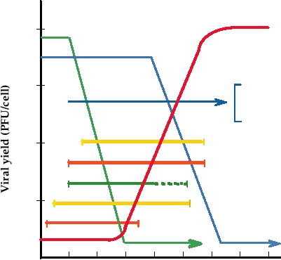
events in the adenovirus life cycle is presented in Fig. 7.21.
gene associated with human tumors. It is both a transcrip-
Virus infection is lytic and the cell eventually dies.
tional activator and repressor. It activates the transcription
of genes whose function is to arrest cell cycle progression.
E1B blocks the activity of p53, and when p53 is inactive or
Adenovirus Oncogenes
absent, cell cycle progression continues. Ad5 E1B binds p53
and sequesters it outside the nucleus, whereas Ad12 E1B
Two early genes of adenovirus, E1A and E1B, encode pro-
does not appear to bind p53 but inhibits its activity in some
teins that induce the cell to enter S phase, in which cellular
indirect way.
DNA is replicated. E1A targets a number of cell proteins that
The net result of the expression of E1A and E1B is the con-
are involved in cell cycling, forming complexes with Rb, p107,
tinued cycling of the cell. The expression of these two genes
p130, p300, and several other cellular proteins. These proteins
alone will transform cells in culture. Rat cells transformed by
are listed in Table 7.11 for two forms of E1A that result from
Ad12 (HAdV-A) will produce tumors in syngeneic newborn
differential splicing, called 12S E1A (because it is translated
104
Infectious Virus
Host DNA Synthesis
Host Protein Synthesis
103
Interference with host defenses
(antagonize IFN-α, IFN-β, TNF-α;
antagonize CTL recognition)
Virion protein synthesis (L1-L5)
102
Late transcription
Viral DNA replication
101
Early protein synthesis
Early transcription (E1A, E2A, E4)
5
10
15
20
25
30
35
40
Hours post infection
FIGURE 7.21 Relative timing of major events during the adenovirus life cycle. Example shown is for HeLa cells
infected with adenovirus at a multiplicity of infection (MOI) of 10. L1L5 are the major late transcription units mapped
in Fig. 7.20. Adapted from Fields et al. (1996), p. 2119 and Ginzberg (1984), Fig. 1 on p. 2.
TABLE 7.11 Cellular Proteins That Bind Directly
A.
Adenovirus
to Adenovirus E1A Proteinsa
p300
Binds tob
E1A binding
Other proteins
E1B
protein
12S E1A 13S E1A
in complex
p53
E1A
p107
pRB
+
+
None known
Rb
p107
+
+
Cyclin A, cdk2/cyclin E, cdk2
p130
+
+
Cyclin A, cdk2/cyclin E, cdk2
p300
+
+
None known
-
TATA
+
TBP-associated factors
B.
SV40
binding
proteins
p300
(TBP)
-
ATF-2
+
None known
Large-T
p53
-
YY1
+
None known
p107
a
Rb
E1A is the first viral transcription unit to be expressed. In the early phase
of infection, differential splicing results in two mRNAs from this gene,
which sediment at 12S and 13S respectively. The translation products of
these two mRNAs are referred to as 12S E1A and 13S E1A.
b
RB is the retinoblastoma susceptibility protein; cdk2 is cyclin-dependent
C.
Papillomavirus
kinase 2; ATF-2 is a member of the ATF family of transcription factors;
YY1 is a human transcriptional repressor. These cellular proteins have
been shown to bind directly to the E1A protein in a biologically relevant
E7
E6
p53
way. See also Fig. 7.22.
Source: Adapted from Fields et al. (1996), p. 2122.
Rb
FIGURE 7.22 Known interactions between the oncogenic proteins
rats but cells transformed by Ad2 or Ad5 (HAdV-C) are not
(shaded with pink patterns) of an adenovirus, a polyomavirus, and a
tumorigenic. The differences in the ability to induce tumors
papillomavirus and cellular proteins that are regulators of cell cycle
appear to be in the immune responses of the host. In par-
progression. (A) Protein E1A of adenoviruses binds to the Rb family,
ticular, Ad12 appears to interfere more effectively with host
promoting entry into S phase. The 19-kD form of E1B also binds to p53,
CTL responses, as described in Chapter 10. Although some
blocking apoptosis. (B) The large T antigen of the polyomavirus, SV40,
interacts with the Rb family of proteins as well as with, p53 (see also Fig.
adenoviruses cause tumors in rats, there is no evidence that
7.28). (C) The human papillomavirus proteins E6 and E7 bind Rb and p53,
they do so in humans. The regulation of cell cycling may be
respectively, the latter promoting the destruction of p53. Adapted from Berg
different in rodents and humans, or the adenovirus proteins
and Singer (1997), Fig. 1.35 on p. 61.
may interact with rodent and human regulatory proteins in
different ways.
effector products of the interferon pathway. VA RNAs are
The proteins targeted by E1A and E1B are key regulatory
highly structured RNAs that are abundantly produced and
elements of the cell. The polyomaviruses and the papilloma-
act as dsRNA decoys. VA RNA binds to PKR, which requires
viruses target many of these same proteins in order to induce
bound dsRNA for activity, but does not activate the enzyme.
cell cycling, emphasizing their importance in the control
VA RNA also binds to several molecules in the RNA inter-
of the cell cycle. The oncogenes of these three families of
ference (RNAi) pathway, including Exportin 5 required for
viruses and their interactions with these key cellular proteins
the export of pre-microRNAs from the nucleus, and DICER
are illustrated schematically in Fig. 7.22.
required for processing of these RNAs, and VA RNA proc-
essed by DICER binds to RISC (see Chapter 10). The result
Interference with Host Defenses
is to saturate this pathway, thereby inactivating it.
Adenoviruses also inhibit the lysis of infected cells by
Adenoviruses interfere with host antiviral defenses in
CTLs. Ad12 E1A protein blocks the transcription of the
multiple ways. This topic is covered in detail in Chapter 10,
genes for class I MHC molecules, whereas the Ad2 or Ad5
so only a summary is presented here. Adenoviruses have two
E3 19-kDa protein prevents the export of class I MHC mol-
independent mechanisms to suppress the interferon system.
ecules to the cell surface. In either case, presentation of
First, E1A inhibits the transcription of interferon response
peptide antigens to CTLs is blocked.
genes by inhibiting the activity of ISGF3, a cellular tran-
Apoptosis is a cell suicide pathway by which cells die
scription factor. Second, VA RNA prevents the activation
after infection by many viruses. Adenoviruses encode
of a protein kinase called PKR, which is one of the major
proteins that delay the advent of apoptosis in order to give
to some extent Ad3 and Ad4, are the adenoviruses most
the virus more time to replicate. First, E3 region proteins
often associated with severe disease, and Ad7 accounts for
interfere with the action of TNF-α, blocking TNF-α-induced
about 20% of adenoviruses reported to the World Health
apoptosis. Second, E1B blocks, or at least delays, apoptosis
Organization.
otherwise induced by E1A.
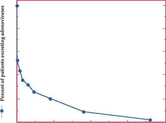
Virus can be shed for months following primary infec-
Because adenoviruses block the antiviral defenses of the
tion, especially in stools (Fig. 7.23). Children who are shed-
host, they have the ability to persist in the infected host for
ding virus are infectious and spread is particularly efficient
considerable periods of time. Virus may be present in tonsils
between close family members. Figure 7.24 diagrams the
and adenoids and may be shed in the stools for a year or
spread of infection within four families in which older sib-
more following primary infection (Fig. 7.23).
lings were excreting adenovirus at the time of the birth of the
youngest child. In all cases the new baby was infected within
a year. Day care centers are also important in the spread of
these viruses.
Adenoviruses and Human Disease
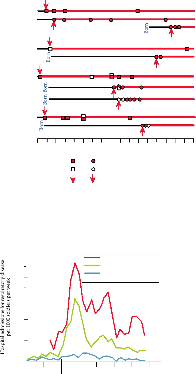
Adenoviruses also cause respiratory disease in adults
and probably account for about 3% of such illnesses. The
The human adenoviruses replicate primarily in the upper
disease is usually mild, but Ad4 and Ad7 have caused
respiratory tract or in the gastrointestinal tract. Some rep-
epidemics of more serious respiratory illness in military
licate well in both while others express a tropism for one
recruits. Such epidemics of acute respiratory disease have
or the other. Spread of the viruses is by a respiratory route
resulted in the infection of 80% of the recruits in a unit and
or by an oralfecal route. Many infections by adenoviruses
2040% of these have required hospitalization. The stress,
appear to be asymptomatic or to result in only mild illness,
crowding, and bringing together of young men from dif-
but about 5% of acute respiratory disease in children under
ferent backgrounds and from all over the country seems
5 years old is due to adenovirus infection. Some serotypes
to potentiate the illness. Illness may be exacerbated if
can also cause gastroenteritis, but the overall importance
infection begins by deep inhalation of aerosolized virus,
of these viruses as causative agents of gastroenteritis is not
which results during the vigorous exercise that is required
resolved. Ad1, 2, and 5 are the most common viruses found
of recruits. Such epidemics result in protective immunity
in human populations, and antibodies to these viruses
because seasoned troops do not suffer recurrent epidemics
are present in about one-half of all children. Ad7, and
100
80
60
40
20
00
200
400
600
800
Days following initial infection
FIGURE 7.23 Adenovirus shedding by patients. The percent of 133 patients who shed virus in stools for at least the
number of days indicated after an initial adenovirus infection is plotted. Note that the last point is almost 2 years. Data
from Strauss (1984).
Family
Age
4 yr
2 yr
A
?
3 yr
B
17 mo
C
recrudescent
2 yr
D
1
2
3
4
5
6
7
8
9
10 11 12 13 14 15 16 17
Months
Male
Female
Fecal shedding
Respiratory shedding
First excretion
FIGURE 7.24
Adenovirus transmission between siblings in four families. Schematic representation of infection with
adenovirus of newborn babies in families where one or more older siblings was already infected. The newborns became
infected between 3 and 12 months after birth. It is believed that shedding of virus was continual (red lines) after the first
virus-positive stool sample from a given child. From Fox et al. (1969).
Special Training Unit (recruits)
Field Artillery (recruits)
Field Artillery Battalions
40
(seasoned men)
30
20
10
Nov
Dec
Jan
Feb
Mar
Apr
May
1944
1945
FIGURE 7.25
Admission to the base hospital for treatment of respiratory disease, presumably primarily adenovirus
related, of two recruit groups and one group of seasoned troops. From Dingle and Langmuir (1968).
Search WWH :