With the development of tail flukes for producing propulsion in whales, manatees (Trichechus spp.), and dugongs (Dugong dugon), the pelves and hind limbs became vestigial structures that now associate only loosely with die spine. The major role of the pelvic apparatus in these forms, when present, is to serve as attachment points for muscles acting on the genitalia and the abdominal body wall. Marine carnivores, which still maintain close ties with the terrestrial environment, have not had such a dramatic reduction in the pelvis and hind limb structures. Both pinnipeds and the sea otter (Enhydra lutris) have united the toes to form flippers. Phocids, which use the hind limbs to generate swimming thrust and which cannot rotate the hind feet under the body while on land, have highly modified hind limbs. Phocid adaptations include increasing the surface areas available for muscles that flex the leg, modification of limb muscles to aid in undulatory movements of the spine, and a general increase in the muscle mass operating on the hind limb (in particular the muscles acting to flex the limb).
I. Cetaceans
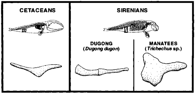
The known fossil record documenting cetacean evolution shows a progressive reduction and loss of hind limb skeletal elements and disassociation of the pelvic girdle from the vertebral column as whales became less dependent on nearshore environments and developed tail flukes to generate swimming thrust. This trend is most marked with the origin of the basilosaurine whales, in which the tibia and fibula became fused with each other and tarsal elements co-ossified into a single immobile mass. Basilosaurines also mark the point during which the pelves became disassociated from the vertebral column. Among modern forms, only vestiges of the hind limb skeleton can be found, and these are contained within the body wall. Mysticetes typically posses only vestigial pelves, whereas the occurrence of hind limb and pelvic elements is more variable among both individuals and species of odontocetes. When present, the pelves bear little resemblance to those of terrestrial mammals and, when undeveloped, may exist only as a band of connective tissue connecting spinal muscles to those of the genitalia and abdominal wall. If present as a bony element, each pelvis is typically cigar or sickle shaped, with only the pelvic bone proper contributing to its structure (Fig. 1). Atavistic femora and occasional tibiae have been described from numerous (mvsticete and odontocete) taxa, although occurrence of these elements is infrequent. Hind limb buds are present during early embrvogenesis of all whale species documented so far, although the mesodermal cells that usually form the internal limb structures die or are reallocated to other functions as limb buds are resorbed later in ontogeny. Retention of a rudimentary pelvis is associated with the attachment of numerous muscles acting on the reproductive organs of both sexes. In males, the pelvis is usually larger relative to that of females. It serves as the site of origin for muscles acting on the genitals and anal region (e.g., the penis retractor and levator ani muscles) and may also serve as a site of attachment for posterior fibers of the rectus abdominis muscle. When present, the pelvis is isolated from the spine (sacral vertebrae are absent) but maintains a soft tissue attachment to the hypaxial spinal musculature. Rearrangements of spinal and pelvic muscles in association with tail-based locomotion have led to considerable controversy over specific identities of muscles in these regions.
II. Sirenians
The evolutionary loss of the hind limb in sirenians closely parallels that of cetaceans (Fig. 1), with modern forms possessing only a vestigial pelvis composed of iliac and ilium bones. In dugongs, each pelvis is long and stick like in appearance, and the ilium and ischium are of subequal length, fusing by 5 years of age in both sexes. In manatees, the pelves are more plate like and cross shaped in lateral view. The ischium is the largest portion of the manatee pelvis, with the ilium forming a small cap on the anterior surface of the bone complex. As in whales, sirenian pelves lack osseous attachment to the vertebral column. In dugongs, the pelves join with the anterior caudal vertebrae by an aponeurosis thought to be homologous with the m. coccygeus, as well as by the retractor ischii and ischio-coccygeus muscles to caudal chevron bones. The pelves serve as the origin for muscles acting on the genital organs (e.g., in females, the constrictor vulvae, constrictor vestibuli, and ure-thralis muscles) as well as some muscles inserting into the skin of the abdominal region [e.g., the transversus abdominis (in part)]. The atavistic appearance of femora has been reported for manatees.
Figure 1 Line drawings of the right pelvis of a cetacean (left) and sirenians (right) in lateral view (anterior toward the right). Position and orientation of the pelvis are indicated by boxes on the skeletal outlines.
III. Marine Carnivores
A. Pinnipeds
The earliest known pinniped, Enaliarctos from the late Oligocene of California, possessed a well-developed hind flipper, and intermediate stages in the anatomical progression from a limb used for terrestrial locomotion to one specialized for swimming are undocumented. Anatomical adaptations of the hind limbs of extant pinnipeds largely reflect strategies adopted by each family for swimming and terrestrial locomotion. Phocid seals and walruses (Odobenus rosmarus) primarily use the hind limbs to generate thrust while swimming and have relatively more muscle mass in the pelvic region relative to the pectoral region and forelimb. Otariids propel diemselves with the forelimbs and have relatively lower pelvic muscle mass. On land, otariids and walruses are able to rotate their hind feet under their body and progress with modified walking motions; phocids lack the ability to rotate their feet under their body and move along the ground with undulatory movements of the body.
Externally, the hind limbs of pinnipeds extend beyond the body contour from the approximate middle or end of the crus. In walruses and phocids the middle digit is the shortest, and digits increase in length both laterally and medially, giving the flipper a crescent shape (more marked in phocids). A thin, extensible interdigital webbing stretches between adjacent digits in these forms. In otariids, the interdigital areas are occupied by thick layers of connective and other tissues, making the hind flipper a much more rigid structure. Emargination of the distal interdigital regions of the flipper confers a scalloped shape to its trailing edge. Claws are reduced in all pinnipeds, although those of the middle three digits tend to be better developed than those of the first and fifth digits. Claws are positioned terminally in phocids and subterminally in walruses, but are located considerably farther proximally in otariids due to the development of distal cartilaginous rods on the ungual phalanges. The presence of these cartilaginous extensions gives the ungual phalanges an hourglass shape and roughened distal ends. The plantar surface of otariid and walrus flippers is hairless, with moderately developed foot pads related to their ambulatory terrestrial locomotion. Foot pads are lacking on phocids. With the exception of interdigital regions, which are hairless or have only sparse hair, the dorsal pedal surface typically has a hair density subequal or slightly lower than that of the body.
Departures of the skeletomuscular anatomy from the condition observed in typical terrestrial carnivores are most prevalent in phocid seals due to their highly modified (undulatory) terrestrial locomotion and specialized hind limb swimming. The iliac region of the phocid pelvis is expanded laterally, particularly among phocines. This confers a mechanical advantage to the gluteus muscle complex, which inserts onto the greater trochanter of the femur and functions to flex the leg against die water. The ischiopubic region of phocid pelves, posterior to the acetabulum, is elongate relative to otariids and the walrus. This increases the surface area available for attachment of the strong muscles acting to medially flex the leg during the power stroke of the swimming cycle (e.g., the adductor, gracilis, gemelli, ob-turatorius, and semimembranosus muscles). The ischial tuberosity, often misidentified as the “ischial spine,” is greatly enlarged in phocids, but undeveloped in otariids and the walrus. It serves as the site of origin for the biceps femoris muscle, which inserts broadly onto the tibia. The orientation and widening of the biceps femoris in phocids indicate that it is primarily responsible for lifting the hind limb off the ground during terrestrial locomotion, as well as medially flexing the limb during swimming. The pinniped femur is short and stout, and the distal condyles are inclined relative to the long axis of the shaft. The fovea capitis of the femoral head is lacking. This indicates the loss of the teres ligament, which normally maintains the femoral head within the acetabulum of the pelves in terrestrial mammals that have weight-bearing hip joints. In phocids, the lesser femoral trochanter is either reduced or absent, and the two muscles typically inserting onto it have undergone major changes from their usual orientation and function: (1) the iliacus muscle inserts onto the more distal femoral epicondylar crest or proximal tibia and (2) the psoas major muscle, arising from the posterior thoracic, lumbar, and sacral vertebrae, inserts onto the medial surface of the ilium and thus aids in lateral undulation of the spine during swimming rather than acting on the limb. Proximally, the tibia and fibula of most pinniped species become fused prior to maturity. The posterior tibial fossa is deep in phocids, reflecting enlargement of the tibialis caudalis muscle, which originates from this region and inserts onto the tarsus and first metatarsal, acting to plantar flex the pes during the swimming power stroke. The tendon of the flexor hallucis longus muscle passes over a posterior projection of the astragalus that is unique to phocids, limiting dorsal flexion of the pes. This, in combination with the elongated iscio-pubis (which limits anteroventral bending of the spine when phocids are on land), limits the ability of phocids to assume a four-legged stance. Additionally, the tibioastragalar joint of phocids is nearly spherical and would unlikely bear the weight of the animal. This is in contrast to the more rigid, hinge-like joint found in other pinnipeds and terrestrial carnivores. Inserting tendons of the large plantar flexing muscles (i.e., the flexor digitorum longus, flexor hallucis longus, and flexor digi-torum superficialis muscles) often combine together in a corn- plex manner at the level of the tarsals. A united tendon of these muscles sends branches out to the digits, although slips extending to the first and fifth digits tend to be larger in phocids. The pedal formula of all pinnipeds is 2-3-3-3-3, the primitive condition for all mammals. In phocids and the walrus, metatarsals and phalanges of the first and fifth digits are more robust than those of the middle three digits. This is associated with hind limb swimming, where both digits may act as leading edges of the flipper during the complex power stroke. Metatarsal-phalangeal and interphalangeal joints tend to be of the tongue-and-groove type in phocids and hinge-like in otariids and the walrus. However, all pinnipeds have reduced troehleation of the metatarsals and phalanges (Fig. 2).
Figure 2 Line drawings of external flipper morphology (row 1), right pelvis (row 11, lateral view, anterior is toward the right), and right hind limb skeleton [row III, anterior (dorsal) view] of representative species of pinnipeds (left; Otariidae, Callorhinus; Phocidae, Monachus; and Odobenidae, Odobenus) and the sea otter (right). Indicated features of the otariid and phocid hind limb are as follows: i, enlarged ischial tuberosity of phocids; ii, presence of a lesser femoral trochanter in otariids and ivalrus (absent or reduced in phocids); iii, posterior projection of the phocid astragalus, over tohich the tendon of the flexor hallucis longus muscle passes; and iv, first ungual phalanx of the otariid pes showing the lack of a claw and distal roughened surface to which cartilage is attached in life.
Available evidence indicates that blood supplying the crus and pes of phocids passes primarily through the external iliac, femoral, and sapheneous arteries. Maintenance of this primitive condition is related to the efficiency of the system for supplying oxygen and nutrients to the heavily used hind limb musculature. In contrast, otariids have adapted the blood vessels such that most of the blood supplying the distal limb regions passes through the internal iliac and gluteal arteries. The passage of blood via this route is believed to enhance heat dissipation. The walrus is intermediate to these conditions, although detailed description of its anatomy is lacking. The presence of a circulatory countercurrent heat exchange system (rete mirabile) in the hind limbs has been reported for several phocid and otariid species. Anatomy of the spinal nerves serving the hind limb of pinnipeds is poorly known. Available evidence suggests that the lumbosacral plexus has shifted posteriorly by one vertebra, being composed of ventral rami arising from the third through fifth lumbar, first through third sacral, and first caudal vertebrae. Division of this plexus into lumbar and sacral plexi is not possible.
B. Polar Bears and Sea Otters
Adequate descriptions of polar bear (Ursus maritimus) pelvic and hind limb anatomy have not yet been made, but there is little indication that the morphology of this species has diverged appreciably from that of other species of Ursus. Departures of sea otter hind limb anatomy from that of other mustelids, however, are seen more readily. Externally, the leg is enclosed within the loose body skin to the approximate level of the anlde. The digits are bound together by interdigital webbing, although the fourth and fifth digits are bound more closely together than other adjacent digital pairs. The sea otter is unusual in that in overall length the digits decrease in size from the fifth to the first: V > IV > III > II > I. While swimming, sea otters use the hind feet to generate thrust and sweep the leg through the water such that the fifth digit forms the leading edge of the pes. The hair densities for the ankle and interdigital webbing have been estimated at 107,000 and 3300 hairs/cm1″, respectively, compared to a density of 125,000 hairs/cm” for the back. Pads are present on the phalangeal portion of each toe and are variably found ventral to the metatarsals. As with pinnipeds, the fovea capitis is absent from the femur, marking the absence of the teres ligament. The biceps femoris muscle inserts onto the middle of the tibia and maintains the leg in a posterior position. The flexor digit V muscle is very large in the sea otter (relative to other mustelids). This enlargement corresponds to the use of the lateral surface of the pes to lead during the power stroke of the limb. The remaining hind limb anatomy of the sea otter corresponds well with that of terrestrial mustelids (Fig. 2).

![Line drawings of external flipper morphology (row 1), right pelvis (row 11, lateral view, anterior is toward the right), and right hind limb skeleton [row III, anterior (dorsal) view] of representative species of pinnipeds (left; Otariidae, Callorhinus; Phocidae, Monachus; and Odobenidae, Odobenus) and the sea otter (right). Indicated features of the otariid and phocid hind limb are as follows: i, enlarged ischial tuberosity of phocids; ii, presence of a lesser femoral trochanter in otariids and ivalrus (absent or reduced in phocids); iii, posterior projection of the phocid astragalus, over tohich the tendon of the flexor hallucis longus muscle passes; and iv, first ungual phalanx of the otariid pes showing the lack of a claw and distal roughened surface to which cartilage is attached in life. Line drawings of external flipper morphology (row 1), right pelvis (row 11, lateral view, anterior is toward the right), and right hind limb skeleton [row III, anterior (dorsal) view] of representative species of pinnipeds (left; Otariidae, Callorhinus; Phocidae, Monachus; and Odobenidae, Odobenus) and the sea otter (right). Indicated features of the otariid and phocid hind limb are as follows: i, enlarged ischial tuberosity of phocids; ii, presence of a lesser femoral trochanter in otariids and ivalrus (absent or reduced in phocids); iii, posterior projection of the phocid astragalus, over tohich the tendon of the flexor hallucis longus muscle passes; and iv, first ungual phalanx of the otariid pes showing the lack of a claw and distal roughened surface to which cartilage is attached in life.](http://lh4.ggpht.com/_NNjxeW9ewEc/TNGTVJeifsI/AAAAAAAAPX0/yQRQ9eZlyDA/tmpC170_thumb_thumb.jpg?imgmax=800)
