Specialized Terms
coupling A term for processes and structures that convey mechanical events in the environment to the sensitive plasma membrane of a mechanosensory neuron.
encoding The creation of a train of action potentials from a sensory receptor potential.
scolopale A ” sense rod, ” or minute rod-like structure around the distal end of a sensory neuron.
transduction The conversion of mechanical displacement at a nerve cell membrane into a receptor current that changes the membrane potential.
tubular body A microtubule-based structure found in the distal sensory dendrites of many mechanoreceptor neurons.
Mechanoreception is the sense that allows insects to detect their external and internal mechanical environments, including physical orientation, acceleration, vibration, sound, and displacement. The integument and internal organs contain a wide variety of mechanore-ceptors. Prominent receptors, such as surface hairs that mediate touch, or auditory organs, have been studied extensively, but many other physiological functions also depend on mechanosensory signals.
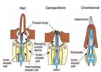
Arthropod mechanoreceptors are divided into two morphological groups: Type I, or cuticular, and Type II, or multipolar. Type I are ciliated receptors, associated with the cuticle, and have their nerve cell bodies in the periphery, close to the sensory endings. They can be subdivided into three major groups (Fig. 1). Hair-like receptors are found on the outer surface in a variety of shapes and sizes, from long, thin hairs to short pegs and scales. A sensory neuron is closely apposed to the base of the hair, and its dendrite contains microtu-bules ending in a structure called the tubular body. It is assumed that movement of the hair compresses the ending, with the tubular body perhaps providing a rigid structure against which the compression can work. Hair receptors can contain additional sensory neurons, such as chemoreceptor neurons in taste hairs. Campaniform (bell-shaped) sensilla are also found on the outer surface, particularly in compact groups near the joints, where they detect stress in the cuticle. Stress moves the bell inward, compressing the dendritic tip containing the tubular body. Chordotonal receptors are generally found further beneath the integument, although they can be connected to the integument by attachment structures. They serve several functions, including hearing and joint movement detection. They often lack tubular bodies but all have dense scolopales surrounding the dendrites and often have multiple mechanosensory neurons.
Type II mechanoreceptors are nonciliated neurons, whose central cell bodies have many fine dendritic endings, each of which is apparently mechanosensitive but lacks the detailed structures seen in Type I receptors. Type II receptors are found in many internal

FIGURE 1 The three major groups of insect cuticular mechanore-ceptors. The receptor lymph space surrounding the sensory ending is formed by a layer of sheath cells and epithelial cells connected by tight junctions. The numbers of sheath cells and their nomenclatures are both variable (see text).
structures, predominantly associated with mesodermal tissues, including the musculature, where they detect muscle tension.
Studies of mechanoreceptor morphology have used many techniques, including light microscopy, scanning and transmission electron microscopy, and immunohistochemistry. Receptor electrophysiology has been studied by three basic methods: (1) Extracellular recordings observe the receptor currents flowing along the axon. (2) Epithelial recordings measure the current flowing through the relatively low resistance of the thin socket tissue or through a cut hair. (3) Intracellular recordings give direct measurements of membrane potentials and currents. Important evidence applicable to insect mechanoreceptors has been obtained from similar structures in arachnids and crustaceans.
Mechanosensation is commonly viewed as a three-stage process in which a mechanical event is first coupled to the receptor cell membrane by mechanical structures, then transduced into a receptor current at the cell membrane, and finally encoded into action potentials for transmission of information to the central nervous system (CNS).
DEVELOPMENT OF MECHANORECEPTORS
Type I sensory neurons are surrounded by specialized sheath cells of varying numbers and names, although the terms trichogen (hair-forming) and tormogen (sheath-forming) are commonly used for the innermost two layers of sheath cells. Development of these cells has been well characterized in several species, but especially in Drosophila external bristles, where many of the genes involved have been identified. A single sensory organ precursor cell divides to give two different secondary precursors, IIA and IIB. IIA divides to form one trichogen and one tormogen cell. IIB gives rise to the neuron, another sheath cell, and sometimes an additional glial cell. The neuron then forms an axon that grows into the CNS. A variety of other noncellular structures, including sheaths, are also found, particularly in dendritic regions. The development of Type II receptors is less well understood.
MECHANICAL COMPONENTS
Extracellular tissues, often with elaborate structures, surround the sensory cells. These structures modify the spatial and temporal sensitivities of the receptors, and are often designed to interact with the outside environment or other parts of the animal, such as cercal hairs detecting air movements or hair plates detecting joint rotation. External structures usually allow detection of mechanical events at some distance from the sensory cell, but make the displacement at the receptor cell membrane smaller than the original movement. Estimates of this attenuation suggest that threshold movements at the cell membrane leading to sensation are in the range 1-5 nm.
TRANSDUCTION AND ENCODING
Mechanically activated ion channels, probably located at the tips of the sensory dendrites, transduce the mechanical stimulus into a receptor current. These transducer channels are permeable to potassium ions, which are more concentrated in the receptor lymph space. The molecular identity of transducer channels is unknown, but there is evidence for both the transient receptor potential (TRP) and acid-sensitive (ASIC) channel families. Mechanotransduction currents are very sensitive to temperature, with activation energy values of 12-22kcalmol-1.
Current flowing through the channels causes a receptor potential that is encoded into action potentials using several different sodium and potassium currents. Action potentials propagate into the central nervous system along axons in nerve roots of the segmental ganglia. Afferent axons have a size range of 1-20 |im and conduction velocities are typically 1-5ms-1. Information is transmitted from mechanore-ceptor axons into the central nervous system via cholinergic synapses.
CENTRAL, PERIPHERAL, AND HUMORAL
MODULATION
Many mechanoreceptors receive GABAergic inhibitory efferent innervation close to the output synapses of their axon terminals. This presynaptic innervation modulates afferent mechanoreceptor information. Some mechanosensory neurons are also modulated by efferent innervation in the periphery and by circulating chemicals such as biogenic amines, of which octopamine has been most thoroughly studied. Octopamine directly excites spider mechanoreceptors in the periphery, but the evidence is less clear in insects.
The extent and functions of peripheral modulation in insects remain to be seen. It is the latest in a series of surprises about the complexity of insect mechanotransduction, but probably not the last.
