One of the most generally known and oft-repeated facts about insects is that they possess three pairs of legs, one pair each on the prothorax, mesothorax, and metathorax. Indeed, this condition is in the fundamental ground plan of insects and is amply represented in the fossil record. The condition inspired Latreille’s taxon Hexapoda (Greek hexa, six, and poda, foot). Exceptions to the hexapodous condition are found in the apodous, or legless, insects that have secondarily lost their legs, typically as a result of selection for an obligatory parasitic or sedentary existence.
The six-legged condition is derived from an ancestral arrangement in which legs occurred on each body segment and bore the same number of segments, each with an inner and outer articulated branch (endite and exite, respectively). Over more than 500 million years of evolution, the multibranched (multiramous), serially uniform legs became modified in the insectan lineage into the characteristic mouthparts, antennae, thoracic legs, and various abdominal appendages, such as genitalia and cerci, while usually becoming lost on other abdominal segments. Further evolution of the six-legged condition in the insectan lineage has resulted in an enormous diversity of structure and function. This structural and functional diversity of legs, along with evolution of the first pairs of exites into wings, without the loss of legs—a condition unique to insects—undoubtedly has been a key factor in the numerical success of insects and their representation in nearly every habitat on the planet. The exquisite diversity in leg structure plays an important role in the taxonomy and classification of insects.
STRUCTURE
In the classic text topic interpretation, the insectan leg has six well-sclerotized segments, arranged proximal to distal: the coxa, trochanter, femur, tibia, tarsus, and pretarsus. A more fundamental and complete segmentation scheme, based largely on the work of Jarmila Kukalova-Peck and Edward L. Smith, involves 11 segments and facilitates the recognition of homologies in legs and leg-derived appendages (e.g., mouthparts and genitalia) among all arthropods. These segments include the epicoxa (present, e.g., as the wing articulation on the thorax of winged insects), subcoxa (embedded in pleu-ral membrane as the pleuron), coxa, trochanter, prefemur (typically fused either with the trochanter or femur), femur, patella (fused with the tibia), tibia, basitarsus, eutarsus (often subdivided), and pretar-sus. A more modern interpretation of the free thoracic leg of extant insects, beginning with the coxa, therefore, recognizes nine segments, which are the classic six, plus a prefemur, patella, and basitar-sus, the latter three visible in only some taxa. The limb appendage of insects that is least changed from the ancestral arthropod condition is the maxilla of jumping bristletails (Archaeognatha), which retained 10 fully articulated segments, two endites, and two exites.
Each segment in the insectan leg, unless secondarily fused or lost, is independently movable by muscles inserted on its base. Thus, subdivisions of the eutarsus, marked by flexible cuticle but without corresponding internal muscles, are not true segments; these subdivisions are referred to as tarsomeres. The areas of flexion between segments are joints, and the well-sclerotized contact points in the joints are the condyles. The various joints contribute to the mechanical efficiency of the leg. The articulation between the coxa and the body, for example, allows the leg to move forward and rearward, whereas that between the coxa and the trochanter allows the leg to be depressed at the beginning, and lifted at the end, of the backstroke.
Leg joints are of two types. Monocondylic joints have a single point of articulation, somewhat like a ball-and-socket joint, and usually are situated dorsally. They allow considerable freedom of movement and are characteristic of the legs of larval insects. Dicondylic joints have an anterior and a posterior condyle, or a dorsal and ventral condyle in the case of the trochanterofemoral joint. They typically limit movement to that of a hinge. Adult legs usually have dicondylic joints, although the tibiotarsal joint is often monocondylic.
The coxa (plural coxae) is typically short and stout. It is set in a coxal cavity and articulates with the thorax at the coxal process of the pleural sulcus (groove). Quite often, it also articulates with the thoracic trochantin and sternum, somewhat restricting its movement. To withstand the forces of movement, the coxa is strengthened by a ringlike basicostal sulcus that sets off a basal sclerite, the basicox-ite. Internally, the basicostal sulcus is expressed as a ridge, the basi-costa, that provides for muscle attachment. Posterior to the point of articulation, the basicoxite is called the meron and in insects such as adult Neuroptera and Lepidoptera, it can be quite large. In higher Diptera, the meron is detached from the coxa and forms a plate in the mesothoracic pleuron. In some insects, an additional external groove, the coxal sulcus, divides the coxa lengthwise.
The trochanter is small and freely movable in a vertical direction on the coxa, but it is often more or less fused with the prefemur plus femur. In the larvae and adults of numerous fossil insects and in extant Odonata, the trochanter and prefemur are distinct segments.
The femur (plural femora) is usually the largest and strongest segment of the leg. It is typically fused to the prefemur in the legs of most winged insects, but in extant Hymenoptera, the two segments are separated by a deep groove. The size of the femur is related to the mass of tibial extensor muscles within it, varying from a small, thick segment in larval insects to an enormous segment in the hind leg of jumping Orthoptera. The femur often is equipped with spines and other cuticular modifications, especially in predatory insects.
The tibia (plural tibiae) usually is fused to the patella (plural patellae) and is typically long and slender in adult insects. In the legs of Odonata and Ephemeroptera, however, the patella is separated from the tibia by an oblique groove. Proximally, the patella is bent slightly toward the femur, allowing the shaft of the tibia to be flexed against the femur for more locomotory power in insects such as grasshoppers. The tibia often bears spines for grooming or for engaging the substrate to aid locomotion. Many insects also have apical or subapical movable spurs on their tibiae.
The tarsus (plural tarsi) is a fused, secondary segment in holo-metabolous larvae and primitive hexapods such as Protura and some Collembola. In collembolans, the tarsus and tibia are fused into a single tibiotarsus. In most insects, a separate segment, the basitarsus, is present and the eutarsus is subdivided into two to four tarsomeres. The ventral surface of the basitarsus and eutarsus often bears pads called tarsal pulvilli that aid movement on smooth surfaces and are especially well developed in some Orthoptera. Thus, the subdivided, flexible eutarsus, in combination with the sharp, curved claws of the pretarsus, superbly adapts insects for climbing. The basitarsus and eutarsus are well endowed with sensory hairs and chemoreceptors. The ventral surface often has a secretory epithelium that produces a wax, possibly for waterproofing, inhibiting the uptake of undesirable water-soluble compounds, or preventing entrapment in surface films.
The pretarsus, also called the acropod or posttarsus, arises from the distal end of the eutarsus. In the Protura, Collembola, and larvae of many holometabolous insects, the pretarsus is a simple, clawlike segment. Typically, however, the pretarsus of insects bears a pair of hollow claws (ungues) and various sclerites and lobes. A scle-rotized, basal unguitractor plate articulates with the eutarsus into which the plate is partly invaginated. The muscles that flex the claws are inserted on a process of this plate. The claws articulate with the unguifer, a median process at the distal end of the eutarsus. A saclike, hollow lobe, the arolium, arises between the claws. In adult Diptera, other than crane flies, an arolium is absent. Instead, a padlike lobe called the pulvillus (plural pulvilli) arises from an auxiliary plate (aux-ilia) beneath the base of each claw, while an unpaired, lobelike or bristlelike process, the empodium, stems medially from the ungui-tractor plate. The pretarsal pads and lobes have adhesive setae (tenent hairs) that allow the insect to climb and hold onto smooth surfaces.
Variation in structure, and hence function, can be found among the three pairs of legs within an individual, as well as between larvae and adults, between males and females, and among taxa. The thoracic legs of many larval insects are serially uniform or sometimes lacking. The legs of adults often vary in structure among the three pairs, although even adults of some insects (e.g., some female Coccidae, Psychidae, and Strepsiptera) are devoid of legs. The variation among the three pairs of legs in adults often is associated with acquisition of food, courtship, and mating. Among the hemimetabolous groups, many Hemiptera-Heteroptera recover a tarsomere in the final molt.
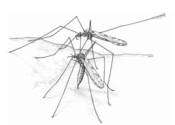
Sexual dimorphism in leg structure is prevalent. The reduced forelegs of nymphalid butterflies have short tarsomeres in females but lack all segments beyond the tibia in males. The forelegs of male Ephemeroptera are typically elongated and used to grasp the female. The slender, elongate legs of crane flies are even longer in males than in females for species in which a guarding male stands over the female during oviposition (Fig. 1); in some crane flies the distalmost tarsomere is prehensile in males and used for holding females. The hind femora of the males of many Coreidae are enlarged for intrasexual fighting. The pulvilli beneath the claws of some flies, such as Tachinidae, are considerably larger in males than in females.

FIGURE 1 Long legs allow male crane flies (Dactylolabis montana) to stand guard over ovipositing females they have recently inseminated.
FUNCTION
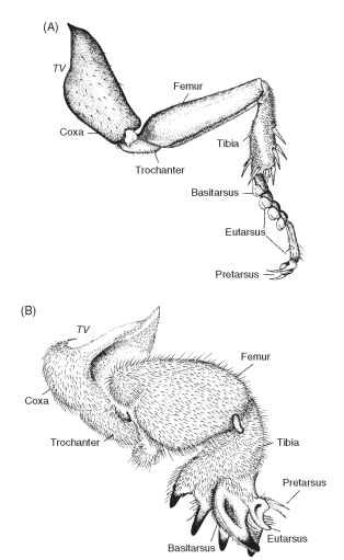
The majority of insectan legs are either elongate, slender, and designed for walking and climbing or cursorial, that is, adapted for running, as in the cockroach (Fig. 2A). During walking, the legs form alternating triangles of support, with the fore and hind legs of one side and the middle leg of the opposite side contacting the substrate as the other three legs move forward. Various modifications allow the legs to be used in other forms of locomotion. Enlarged hind legs of many Orthoptera, fleas, and other insects are saltatorial, meaning they are designed for jumping. The jump of insects such as fleas is aided by a rubberlike protein called resilin in the cuticle that stores and subsequently releases energy for the jump. Powerful, spadelike forelegs of mole crickets, scarab beetles, burrowing mayflies, and other insects are fossorial, or adapted for digging and rapid burrowing (Fig. 2B). Flattened, fringed legs of aquatic insects such as dytiscid and gyrinid beetles and noto-nectid backswimmers serve as oars for paddling or swimming (natatorial legs), while long legs with hydrophobic tarsal hairs and anteapical claws, as in water striders (Gerridae), are for skating on the surface of water. The legs of some insects, although well developed, have lost their locomotory function. The spiny legs of Odonata, for example, are designed for perching or seizing and holding prey captured while the dragonfly is in flight; the legs are ineffectual for walking.
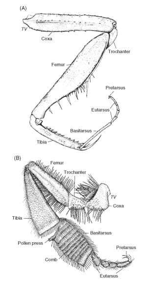
Although typically regarded as agents of locomotion, the legs also have assumed additional, or altogether different, functions. Often, function can be inferred from structure; for example, the thickened, spinose legs of many insects signify a predacious mode of life. Raptorial legs (Fig. 3A), that is, those designed to seize prey, have arisen independently in many insectan lineages. Any of the three pairs of thoracic legs can be raptorial, but the trait is most often expressed in the forelegs (e.g., in Mantodea and Reduviidae) and less frequently in the middle legs (e.g., in some Empididae) and hind legs (e.g., in some Mecoptera).
Legs also play a defensive role, aiding escape by running, jumping, burrowing, and swimming, as well as by kicking and slashing. The spines on the legs of many insects deter predators and competitors and can inflict considerable damage. Insects such as stink bugs and treehoppers deliver powerful kicks at parasitoids and predators that attempt to attack their young. Autotomy, or the loss of legs at predetermined points of weakness, often at the level of the

FIGURE 2 (A) Cursorial foreleg of the Madeira cockroach (Leucophaea maderae). (B) Fossorial foreleg of the northern mole cricket
trochanter, occurs in insects such as crane flies, leaving a predator with only a leg in its clutches as the insect escapes. Legs, whether lost through autotomy or accident, can be regenerated to various degrees in some insects, most famously in walking sticks, if one or more molts follow the amputation.
All legs are equipped with an extensive arrangement of sensory structures that allow the insect to feel, hear, and taste. Chemoreceptors, which are especially prevalent on the basitarsus and eutarsus, provide sensory input on environmental substances and can be used to determine the acceptability of food, ovipositional substrates, and perhaps mates. Mechanoreceptors, most often in the form of hair organs, but also campaniform, chordotonal, and plate organs, provide sensory information on position, movement, and vibrations borne by air and substrate.
In many insects, the legs are used in sound production. Familiar examples include the shorthorned grasshoppers, which have a stridulatory mechanism on the hind femur, involving a series of pegs—the scraper—that is rubbed across a ridged wing vein. Some larval hydropsychid caddisflies have a scraper on the prothoracic femur that is rubbed against a file on the venter of the head. Legs also can be used to produce sound for intraspecific communication by drumming them against a substrate, as in some Orthoptera.

FIGURE 3 (A) Raptorial foreleg of the Carolina mantid (Stagmomantis carolina).(B) Inner surface of the hind leg of the honey bee (Apis mellifera).
To maintain hygiene, insects spend considerable time grooming their body and appendages. Grooming typically is effected by various leg structures, such as cuticular combs (ctenidia), setal brushes, grooves, and notches. The cleaning setae on the foretibia of certain heteropterans are mirror images of the arrangement of antennal setae. The hind leg of honey bees is specially modified to groom pollen from the plumose hairs of the body (Fig. 3B). Combs on the inner surface of the hind basitarsus remove the pollen from the body hairs and pass it to the pollen press between the tibia and the basitarsus. Closure of the press forces the pollen into the pollen basket (corbicu-lum) on the outer surface of the tibia where the pollen bolus is held in place by rows of hairs. Once in the hive, the honey bee removes the pollen, with the aid of an apical spur on the middle tibia.
Legs often are used to hold onto objects, and they bear the relevant modifications, including enlarged segments to house increased musculature, various spines and setae, and adhesive organs. The grasping function is seen, for example, in the pincerlike, spiny raptorial forelegs of many predacious insects. It also is expressed dramatically in certain sucking lice in which the claw folds against a thumblike, spinose process of the enlarged tibia. Flies that feed on the blood of birds typically have a thumblike lobe at the base of each of their talonlike claws that helps them grasp feather barbules. Grasping is common during mating, and especially the males of many insects have legs designed to secure and hold their mates. Adhesion to objects such as mates and prey can be achieved with suction disks on the legs. Male dytiscid beetles have a flattened, disklike arrangement on each foreleg that is formed of the basitarsus and the succeeding two tarsomeres; all three structures bear minute suction cups ventrally that can be applied to the elytra of the female.
The colors and patterns of legs vary from subtle to stark, although their function is often poorly understood. Long-legged insects such as phantom crane flies (Ptychopteridae) and some mosquitoes have banded legs that might render the insect less conspicuous through disruptive coloration. Other configurations of pattern and color play a role in camouflage, courtship, and mimicry. In flies such as some Syrphidae and Micropezidae, the forelegs resemble the antennae of aculeate Hymenoptera, reinforcing the remarkable overall resemblance of fly to wasp.
Other functions ascribed to legs are often highly specialized. In the Embiidina, the basitarsus of each foreleg houses multiple silk glands, and each gland is connected to a seta with an apical pore through which the silk is extruded. The inflated basitarsus of each leg in phantom crane flies contains a tracheal sac, aiding buoyancy during the driftlike flight. Some flies have specialized areas on their legs, particularly on the tibia, that possibly produce pheromones. Insects such as Chironomidae seem to use the legs much as a second set of antennae. In Protura, which lack antennae, the forelegs probably have assumed an antennal (i.e., sensory) function.
Acknowledgments
I thank T. S. Vshivkova for illustrating Figs. 2 and 3, and J. Kukalova-Peck for insightful comments on the manuscript and access to her prepublished work.
