fMHV
RL Plasmid
T7
1 HE
RL
1b
1a
2A
4ab/5
HE
3 NTR
5 NTR
An
CAP
felineS
murineS E
M
EM N
N
Transcribe RNA with T7
3 NTR
5 NTR
Infect feline cells
Electroporate RNA into
infected cells
Incubate 24 hours at 37 C
Plate cells onto monolayer of murine cells
(selects for recombinants)
Plaque purify
1a
1b
2A HE
RL
CAP
An
M
N
murineS E
Full-length attenuated, replication competent MHV
expressing Renilla luciferase (RL)
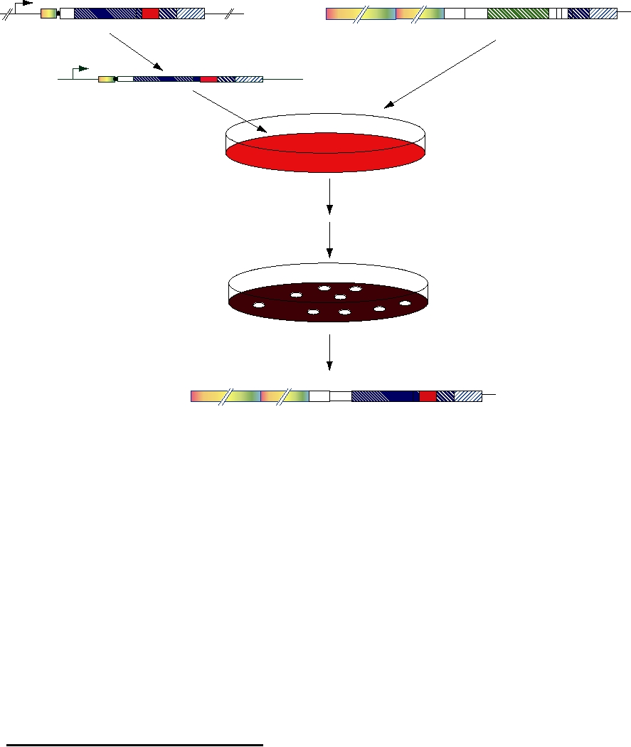
FIGURE 11.6 Construction of a coronavirus expression plasmid. An attenuated and nonreplicating plasmid from
MHV is constructed in which the 5′ end of the ORF 1 sequence is fused to the last 28 codons of HE. In addition, all of the
accessory protein genes are deleted, and the gene for Renilla luciferase (RL) inserted. RNA is transcribed in vitro from
this plasmid using T7 polymerase and the RNA electroporated onto feline cells which have been infected with MHV virus
in which the spike S protein has been mutated to recognize receptors on feline cells (fMHV). After 24 hours these cells
are plated out onto a monolayer of murine cells to select for coronaviruses that have undergone recombination between
the plasmid and the helper virus. Recombinants are plaque-purified and stocks grown.
are unable to infect the cells normally infectable by VSV
several such vaccines will be licensed in the near future.
because they lack the G protein. However, because they con-
There is also expectation that viruses will be useful as vec-
tain the HIV receptor and coreceptor on their surface, they
tors for gene therapy, and numerous clinical trials are taking
do infect cells that express the HIV glycoprotein on their sur-
place. The results to date have been disappointing, but the
face, such as cells infected by HIV. Since VSV is a lytic virus,
promise remains.
the HIV-infected cells are killed.
Expression of Proteins in Cultured Cells
USE OF VIRUSES AS EXPRESSION VECTORS
The use of viruses to express foreign genes in cultured
cells is well established and only a few examples are cited to
Viruses have been widely used as vectors to express a
illustrate the range and purpose of such use.
variety of genes in cultured cells. This use is of long stand-
Hepatitis C virus (HCV) does not grow in cultured
ing and has led to important results. Of perhaps more inter-
cells to titers sufficient to allow studies on the expres-
est are efforts to develop viruses as vectors for medical
sion of viral proteins. The only experimental model for
purposes. The manipulation of virus genomes to develop
the virus is the chimpanzee, which severely restricts the
new vaccines is very promising. Although no licensed
number and nature of experiments that can be done. Thus,
human vaccines have been introduced using this technol-
most of what we know about the expression of the HCV
ogy, clinical trials are ongoing and it is to be expected that
genome has been obtained through expression of parts of
A. Rhabdovirus genome organization
B
5
N
P
M
G
L
tr
le
vRNA ()
3'
T7
vcDNA
Vaccinia expressing T7 polymerase
. Transfect/Infect
T7
T7
VSV
vcDNA
T7
T7
Plasmids with
pL
T7 promoters
T7
pN
encoding
NUCLEUS
pP
VSV P, N, and L
C. Expression of VSV replicase proteins (L, P) and N protein under the control of T7 promoter
D. Transcription of VSV vRNA from cDNA by T7 polymerase; replication of vcRNA,
vRNA, and transcription of VSV mRNAs by VSV replicase/transcriptase
E. ranslation of viral proteins and assembly of infectious virus.
Expressed Products:
T7 RNA polymerase
T7 RNA polymerase
vcRNP (+)
(L, P, N proteins)
VSV G protein
vRNP
mRNAs
An
An
An
An
VSV L protein
N,P proteins
M protein
L
Glycoproteins
VSV N, P proteins
M protein
Infectious VSV
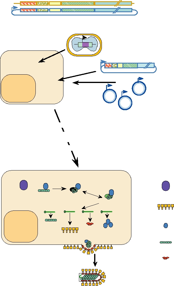
FIGURE 11.7 Rescuing infectious VSV virions from cDNA clones. (A) The rhabdovirus genome organization, and a
schematic of a cDNA clone containing the genome sequence (cDNA copy of vRNA). (B) A susceptible cell is infected
with vaccinia virus expressing the T7 RNA polymerase, and transfected with four separate plasmids: the genome plasmid
from which plus strand anti-genome RNA is transcribed, and 3 individual plasmids expressing VSV N protein, P protein,
and L protein, all under the control of T7 promoters. (C), (D), (E) Steps in the synthesis of infectious virus are described
within the figure. Infectious virions bud from the cell and can infect a new susceptible cell. Adapted from Conzelmann
and Meyers (1996).
A. Genome organization of plasmid with CD4 and CXCR4 in place of VSV G protein
le
N
P
L
tr
CD4 CXCR4
M
Mutant vRNA (-)
5'
3'
T7
Mutant vcDNA
B. Transfect/infect
Vaccinia expressing T7 Polymerase
T
7
T7
Mutant vcDNA
T7
Plasmids with
T7
pL
T7 promoters encoding
T7
pN
VSV P, N, and L
NUCLEUS
pP
Expressed Products:
C. Expression, transcription, translation, replication
T7 RNA polymerase
T7 RNA polymerase
vcRNP (+)
CD4 Protein
(L, P, N proteins)
vRNP
CXCR4 Protein
mRNAs
An
An
An
An
An
VSV L Protein
N,P proteins
M protein
RT
CD4 CXCR4
VSV N, P Proteins
M protein
D. Infect new cell
HIV-infected cell
Infectious VSV
Eukaryotic Cell
With CD4,CXCR4
Becomes infected
Susceptible to VSV
(Mutant VSV)
with
Not infectible
Mutant VSV
with
Mutant VSV
HIV-infected cell
dies
FIGURE 11.8 Producing a mutant VSV targeted to kill HIV-infected cells. (A) Genome of a rhabdovirus in which
the glycoprotein G gene has been replaced with sequences encoding CD4 and CXCR4, the HIV primary receptor and
coreceptor, and a schematic of a cDNA clone containing the genome sequence (cDNA copy of vRNA). (B) A susceptible
cell is infected with vaccinia virus expressing the T7 RNA polymerase, and four separate plasmids: the mutant genome
plasmid from which full-length vc (plus strand) RNA is transcribed, and three individual plasmids expressing VSV N
protein, P protein, and L protein, all under the control of T7 promoters. (C) Plus-strand mutant vcRNA is transcribed
and encapsidated with N, P, and L. The RNP then replicates and both viral proteins and CD4 and CXCR4 are expressed
from individual mRNAs transcribed from genome sense RNPs. Virions bud from the cell and (D) cannot infect a new
susceptible cell as before in Fig. 11.7, but can infect an HIV-infected cell expressing HIV env proteins on its surface.
Infection with VSV is cytolytic and the HIV-infected cell dies. Adapted from Conzelmann and Meyers (1996).
the genome by virus vectors, often by recombinant vac-
Viruses as Vectors to Elicit an
cinia virus. These studies have resulted in an understand-
Immune Response
ing of the two viral proteases within the HCV genome, the
Much effort is being put into the development of viruses
processing pathway through which the polyprotein trans-
as agents to immunize against other infectious agents,
lated from the genome is processed, the function of the
including other viruses. Such an approach has a number of
viral IRES, and the function of the viral replicase, among
advantages. There is a large body of experience in the use of
other results. The use of virus vectors means that such
attenuated or avirulent viruses as vaccines. Many of these,
studies on HCV can be conveniently conducted in mam-
such as vaccinia virus or the yellow fever 17D virus, both of
malian cells under conditions that are related to the natural
which have been used to immunize many millions of peo-
growth cycle of the virus.
ple, can be potentially developed as vectors to express other
Norwalk virus is another virus for which there is no cell
antigens, such as those of HCV or HIV. Use of a live virus
culture system. The virus can be grown only in human vol-
as a vector to express antigens of other pathogens has many
unteers, again limiting the range of studies that can be done.
of the advantages of live virus vaccines. This includes the
Virus particles isolated from the stools of infected volunteers
fact that only low initial doses are required, and therefore the
are often degraded and difficult to purify to homogeneity.
expense of vaccine production may be less; that subsequent
Thus, structural studies of infectious virus have been limited.
virus replication leads to the expression of large amounts of
Expression of cDNA copies of the structural proteins of the
the antigen over an extended period of time, and the antigen
virus in baculovirus vectors has allowed the production of
folds in a more or less native conformation; and that a full
large amounts of viral structural proteins that spontaneously
range of immunity, including production of CTLs as well as
assemble into virus-like particles. These virus-like particles
of humoral immunity, usually develops.
have been studied by cryoelectron microscopy, and detailed
No human vaccines have been licensed that use such
information on the structure of the virus has been obtained
recombinant viruses, but there are ongoing clinical trials of
in this way.
several potential vaccines. Several trials of candidate vaccines
Baculoviruses are also widely used to prepare large
against HIV have been conducted that use vaccinia virus or
amounts of protein for crystallographic studies. Such studies
retrovirus vectors to express the HIV surface glycoprotein.
require 20 mg or more of protein, and the baculovirus system
These trials have been moderately successful in the sense that
can be used to prepare such quantities. An advantage of the
immune responses to HIV glycoprotein were obtained, but
system is that the protein is made in a eukaryotic cell, which
these immune responses were not particularly vigorous and
can be important for obtaining the protein folded into its cor-
it is not known if the immune response is protective. HIV is
rect three-dimensional conformation. Also of importance is
able to persist in infected patients despite a vigorous immune
the use of secretion sequences in the constructs that lead to
response, and sterilizing immunity might be required. Further,
the secretion of the protein from the infected cell, making it
the HIV surface glycoprotein is highly glycosylated and neu-
easier to purify the protein.
tralizing antibodies are difficult to obtain. Studies in mon-
Even for viruses for which cell culture systems exist,
keys with related vaccines against simian immunodeficiency
the use of virus vectors that express to higher levels can be
virus have given mixed results. In most such trials, immune
advantageous. There are cell culture systems in which rubella
responses were generated, but these were not fully protective.
virus will grow and plaque, and there is a full-length cDNA
One recent trial did generate a protective response, however,
clone of rubella virus from which infectious RNA can be
giving hope that continued efforts in this direction will ulti-
recovered. However, the cell culture systems produce only
mately work out. Recent studies with anti-HIV drugs given
low amounts of virus proteins, especially of the nonstructural
very soon after infection found that limiting the replication of
proteins, and it has been difficult to study the expression and
the virus early appears to allow the generation of a protective
processing of the nonstructural polyprotein. Expression of
immune response in at least some patients. Although such
the nonstructural region of rubella virus in vaccinia virus
studies remain preliminary, they do suggest that a nonsteri-
vectors or in Sindbis virus vectors has allowed the produc-
lizing immune response that restricts virus replication early
tion of much larger quantities of the polyprotein precursor.
might prove to be protective.
This has been used to determine the processing pathways,
Other clinical trials have also tested poxviruses as vec-
the identification of the virus nonstructural protease, and the
tors. Vaccinia virus has been used in an attempt to immunize
identification of the cleavage sites that are cleaved by this
against EpsteinBarr virus, and canarypox virus has been used
protease.
as a vector for potential immunization against rabies virus.
As a final example, vaccinia virus vectors and Sindbis
Although no licensed human vaccines use poxvirus vectors,
virus vectors have been used to map T-cell epitopes for
veterinary vaccines that are based on poxvirus vectors are in
a number of viruses (Chapter 10). For this, defined regions
use. One such vaccine consists of vaccinia virus that expresses
of a viral protein are expressed in order to determine whether
the rabies surface glycoprotein. This vaccine has been used
a particular T-cell epitope lies within that region.
to immunize wildlife. The recombinant vaccinia viruses are
number of deaths and neurological sequelae in people that
spread in baits that are eaten by wild animals that serve as
survive the encephalitis (Chapter 3). Vaccines in widespread
reservoirs of the virus, such as skunks, raccoons, foxes, and
use are inactivated virus vaccines, and the difficulties in pre-
coyotes. This approach has been useful in limiting the spread
paring the large amounts of material required and delivering it
of rabies in wildlife populations. Other poxvirus-based vac-
to large segments of the population are significant. An attenu-
cines include vaccinia virus vectors to protect cattle against
ated virus vaccine, SA14-14-2, has been prepared in China by
vesicular stomatitis virus and rinderpest virus, and to immu-
passing the virus in cultured cells and in rodent tissues. This
nize chickens against influenza virus; pigeonpox virus vectors
vaccine is safe but overattenuated, so that the effectiveness
to immunize chickens against Newcastle disease virus; fowl-
is only 80% after a single dose. In contrast, the yellow fever
pox virus vectors to immunize chickens against influenza,
virus (YF) 17D vaccine has an effectiveness of virtually 100%
Newcastle disease, and infectious bursal disease viruses; a
after a single dose, and immunity is long lasting, probably life-
capripox virus vector to immunize pigs against pseudorabies
long. A candidate JE vaccine has been developed that consists
virus; and a canarypox virus vector used to immunize dogs
of the 17D strain of YF virus in which the prM and E genes
against canine distemper virus. Thus, it should be possible to
have been replaced with those of JE, as illustrated in Fig. 11.9.
develop human vaccines based on poxvirus vectors.
Four chimeric viruses were tested. The JE structural proteins
In a quite different approach, clinical trials of a novel
were taken from either the virulent Nakayama strain or from
vaccine against Japanese encephalitis (JE) virus have been
the attenuated SA-14-14-2 strain. In both cases, chimeras
conducted. JE is a scourge in parts of Asia, causing a large
containing all three structural proteins from JE were tested
Original Constructs
Infectious 17D yellow fever cDNA clone
prM
C
E
NS1
NS5
Japanese encephalitis (Nakayama) cDNA (virulent)
Japanese encephalitis (SA) cDNA (human vaccine strain)
Chimeric Constructs
Titer after RNA
PRNT
transfection of anti-YF anti-JE
YF/JE-S prM-E
VERO cells
106
<1.3
3.1
6.3
C
prM
E
NS1
NS5
YF/JE-N prM-E
10 7
<1.3
3.4
2.0
YF/JE-S C prM-E
<10
NA
NA
YF/JE-N CprM-E
<10
NA
NA
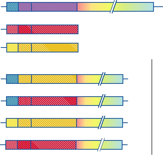
FIGURE 11.9 Construction of yellow fever/Japanese encephalitis chimeric viruses. Starting with the full-length
cDNA clone for 17D yellow fever virus, a number of chimeric viruses were constructed in which the M and E proteins
were replaced with those of different strains of Japanese encephalitis virus. However, when C, M, and E of JE were
put into the yellow fever clone, no viable virus was obtained. Both prME chimeras grew well in tissue culture, and
were neutralized by anti-JE antiserum. YF/JE-S prM-E was attenuated, and did not kill adult mice by intracerebral
inoculation, but YF/JE-N prM-E was neurovirulent. PRNT is the log reciprocal of the dilution yielding 50% plaque
reduction neutralization, based on 100 PFU on LLV-MK cells, using either YF or JE hyperimmune ascitic fluid. Adapted
from Chambers et al. (1999).
Search WWH :


