Embryogenesis is the process by which a larva or a juvenile is built from a single egg. The fertilized egg divides to produce hundreds of cells that grow, move, and differentiate into all the organs and tissues required to form a larva or juvenile. Embryogenesis is extremely diverse in different insect species. For each generalization that can be made about some aspect of insect development, numerous variations and multiple exceptions exist. In some species a single egg gives rise to several thousand larvae, in others, embryos devour their mothers prior to hatching. The most extreme variations are found among insects that parasitize other insects. This article presents a generalized view of some of the more regular features of insect development.
EGG MEMBRANES
Most insects lay eggs in terrestrial environments. For the most part, an insect egg forms a self-reliant developmental system that is generally impervious to the external environment, although sensitive to temperature, which serves as an important cue for many developmental events. Insect eggs are typically quite large, both in absolute dimensions and relative to maternal body size, and well-provisioned with yolk. Eggs vary from about 0.02 to 20 mm in length. To prevent desiccation, they are covered by some of the most resistant and impenetrable egg coverings found in the animal kingdom. Egg contents are protected by a vitelline membrane and covered by an external hard shell, the chorion. The chorion, vitelline membrane, and egg membrane itself surround the internal contents of the fertilized egg: the zygote nucleus and two types of macromolecule. Nutritive material such as yolk proteins, lipids, and carbohydrates are used for nourishment and growth of the embryo. Patterning molecules (such as specialized proteins and mRNAs) direct major events in embryo-genesis including establishment of embryonic polarity, segmentation, and gastrulation.
EGG CLEAVAGE
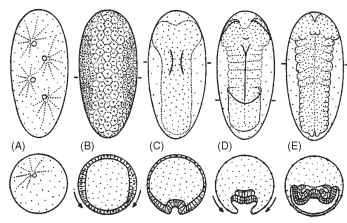
Development in nearly all animals involves a period in which the egg is subdivided into increasingly smaller cells. Compared with other animals, insect eggs undergo an unusual type of cleavage. In most animals, cleavage involves subdivision of both cytoplasm and nuclear material, to form individual cells called blastomeres. In contrast, the early cleavages of most insects involve only nuclear subdivisions (karyokinesis) and are not accompanied by cleavage of the cytoplasm (cytokinesis). This type of cleavage is called syncytial cleavage and results in the formation of a common compartment (syncytium), where up to several thousand nuclei reside (Fig. 1).
It is unclear how syncytial cleavage evolved in insects. The sister group to insects, the entognathans, has both total egg cleavage (Collembola) and syncytial cleavage (Diplura). Basal arthropods such as chelicerates, which include horseshoe crabs, spiders, and scorpions, have both syncytial and total cleavages. Even Onychophora,which are believed to be a sister group to Arthropoda, exhibit both syncytial cleavage (in oviparous species with yolky eggs) and total cleavage (in placental, viviparous species). However, it is reasonable to believe that syncytial cleavage in arthropods and insects evolved from total cleavage in the ancestor to the arthropods.
BLASTODERM FORMATION
In most insects, the early syncytial cleavages proceed rapidly to form a syncytium with up 6000 nuclei. Syncytial nuclei are surrounded by islands of cytoplasm that separate the nuclei from one another. In general, these early cleavages are synchronized and, as the nuclei divide, they separate, such that there is regular spacing between them. Nuclei and associated cytoplasm are referred to as energids. Upon reaching a critical density, the energids migrate to the periphery of the egg. The arrival of the energids at the surface is sometimes visually apparent as bumps along the surface. At the periphery, the energids continue to undergo several rounds of division. In most insects, some of the nuclei remain in the yolk mass. These nuclei subsequently cellularize and become vitellophages, which serve to break down yolk to be used for embryo nutrition. Following the arrival of energids at the egg periphery, the egg membrane invaginates from the egg surface to surround each of the individual syncytial nuclei, marking the end of the syncytial stage of development. The single sheet of cells thus formed at the periphery of the egg is the cellular blastoderm (Fig. 1 ).
FORMATION OF GERM CELLS
In some species, distinctive granular inclusions can be found in the posterior cytoplasm of the egg. The cells that inherit these granules become the germ cells and eventually migrate into the ovaries or testes to become sperm and eggs. When the germ cells are

FIGURE 1 Diagram of the basic pattern of early insect embryo-genesis: ventral views of eggs, anterior poles at top, are shown above cross sections at the levels indicated by bars in top row. (A) Syncytial cleavage. (B) Formation of the cellular blastoderm: arrows show that the lateral cells are coalescing toward the ventral surface to form the germ anlage. (C) Gastrulation. The prospective mesoderm begins invagination along the midline of the germ anlage. (D) Germ band after gastrulation, with segment borders (dotted) and amniotic folds forming: arrows indicate the movement of the serosal cells to enclose and cover the developing germ band. (E) Advanced germ band stage, with appendage buds, and transient coelomic sacs formed by the mesoderm.
ablated, the germline is missing and the individual is sterile, as noted in 1911 by Hegner. The nuclear energids (in some species, such as Drosophila) arrive at the posterior before reaching any other egg region, and the cells that will include this specialized cytoplasm cel-lularize earlier than any of the other cells. Germ cells rarely grow or divide during embryogenesis. The early segregation of these cells is thought to protect them from potential errors incurred during division and differentiation that might damage the genetic material necessary to build the next generation. In other species (e.g., most Lepidoptera), segregation of the germline occurs in the middle of the blastoderm; in other species no apparent germ cells can be detected at the blastoderm stage.
SEROSAL FORMATION
Only blastoderm cells destined to form the embryo coalesce to form the germ anlage, which later develops into the germ band. The cells that do not contribute to the germ anlage form an extraembry-onic membrane called the serosa. In most species, the boundary between the future serosa and the future embryo ruptures, and the serosal cells migrate over and envelope the embryonic primordium and yolk cells (Fig. 1). However, there is variation in how the serosa is formed. In extreme cases like dipterans, the serosa cells do not migrate over the germ anlage but remain as a cluster of cells on one side of the egg. In addition to the serosa, a second protective membrane, the amnion, forms later from the cells immediately adjacent to the germ anlage. These cells proliferate, flatten, and elongate. As they extend over the germ band, they resemble a sleeping bag being pulled up from the posterior and down from the presumptive head lobes of the germ band. Ultimately, the amnion cells meet in the middle of the embryo and form a single cell layer that lies between the embryo and the now separate serosa. In some derived, holom-etabolous species (e.g., Drosophila), this membrane has become vestigial, and the cells never migrate over the germ band.
GERM ANLAGE FORMATION
The size of the germ anlage varies relative to the length of the egg. In nearly all species, the nuclei arrive at the periphery to form a blastoderm that encompasses the whole surface of the egg. In metamorphic species, such as fruit flies and honey bees, the germ anlage forms from nearly the entire blastoderm surface. However, in direct-developing species (such as the grasshopper and cricket), after the formation of a uniform synctyial blastoderm, nuclei migrate and aggregate near the posterior pole, where the germ anlage forms. The germ anlage thus forms from a relatively small proportion of the blastoderm. In the former case, called long-germ-type embryos, the complete body pattern (head, gnathal, thoracic, and abdominal segments) is patterned at the blastoderm stage and all segments appear nearly simultaneously in development. In contrast, in short-germ-type embryos, the head lobes, the most anterior trunk segments, and the posterior terminus are patterned first. Additional segments are added progressively, through proliferative growth. Some insects develop with germ types intermediate between these two extremes. The pleisiomorphic condition for insects is believed to be an intermediate-sized germ anlage. Short/intermediate germ embryogenesis is predominant in direct-developing hemimetabolous insects; more derived, metamorphic insects exhibit long-germ development. However, this division is not clear-cut. In some insect families, closely related species can exhibit both short- and long-germ types of development.
GASTRULATION
Formation of the cellular blastoderm is followed by gastrulation, the process of cellular invagination that results in the formation of a layered embryo comprising two germ layers. Cells that remain at the blastoderm periphery will form the ectoderm, and cells that invagi-nate below the ectoderm will form the mesoderm. The presumptive mesoderm in most species consists of a strip of cells along the ventral midline (Fig. 1). Gastrulation can happen in any number of ways: by the mesodermal cells invaginating simultaneously, as in Drosophila, or by cells invaginating sequentially, beginning at the anterior, while the gastrulation furrow progresses toward the posterior. Most often the presumptive mesoderm lies along the ventral midline, but in the apterygote thysanuran Thermobia domestica, cells migrate inward from every part of the germ band. Regardless of the mechanism of gastrulation, the end result is a bilayered embryo, with mesodermal precursors underlying the ectoderm.
SEGMENTATION
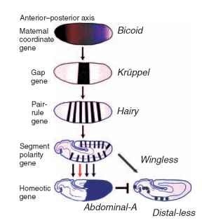
Segmentation refers to the process by which repeated units of similar groups of cells, the metameres, are created. Segmentation proceeds nearly simultaneously with gastrulation. Current understanding of the process of segmentation comes from the genetic dissection of development in Drosophila by Ed Lewis, Christiane Niisslein-Volhard, and Eric Wieschaus. These researchers used a large-scale mutant screen to uncover developmental defects in Drosophila. They found a complex genetic regulatory cascade that specifies the insect body plan (Fig. 2), which has since been shown to have many commonalties with molecular patterning in vertebrate embryos. The three were awarded the Nobel Prize for their efforts. However, even before the genetic dissection of segmentation, the

FIGURE 2 Simplified diagram of the segmentation gene cascade in Drosophila melanogaster and its relation to limb development. Diagrammatic representation of some of regulatory interactions between genes in the Drosophila segmentation cascade: a maternal coordinate gene, a representative gap gene, a pair-rule gene, the segment polarity gene, a homeotic gene, and the limb-patterning gene Distal-less.
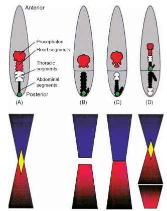
elegant work of Klaus Sander had defined the basic mechanisms of embryo patterning that helped in the interpretation of the new genetic data. By analyzing the outcomes of embryonic manipulations ofleafhopper embryos (Eucelis), Sander concluded that two morphoge-netic gradients specify the pattern elements along the anteroposterior axis of the germ band: one gradient with a high point in the posterior pole and another with a high point at the anterior pole (Fig. 3 ). In 1976 Sander reviewed these experiments, and a large body of other experimental work on insect embryos, setting the stage for modern understanding of the molecular basis of insect development.
MOLECULAR CIRCUITS THAT REGULATE SEGMENTATION
In the cascade of gene activity that generates the segmental pattern of the embryo (Fig. 2) , segmentation proceeds by a progressive refinement of positional information that will eventually specify groups of cells that form metameric units. Refinement is initiated with maternally provided proteins that form gradients from the anterior to the posterior of the egg and early cleavage stages. These

FIGURE 3 Double gradients. (A) Leafhopper embryo consists of head (red), thoracic (white), and abdominal segments (black). Posterior pole of the egg is marked by bacterial symbiont (green). (B) After early egg ligation, anterior and posterior fragments form fewer segments than in the normal embryo. (C) Late ligations result in more segments formed in anterior and posterior fragments. (D) Finally, when posterior material has been displaced anteriorly and the egg ligated just below the symbiont marker a mirror-image duplication was formed. Schematic of corresponding anterior (blue) and posterior (red) gradients and their overlap (yellow) below images illustrates possible anterior and posterior gradients and their interactions in each experimental intervention.
gradients of maternal proteins provide the coordinates that position the front and back of the embryo; hence the genes that encode these proteins are called the maternal coordinate genes. The function of these maternal gradients is to activate the gap genes, which are so named because when their function is lacking, the segmental pattern of the embryo has large gaps in it (e.g., the three thoracic segments may be missing, or the first few abdominal segments.
The proteins encoded by the gap genes in turn activate the pair-rule genes. The pair-rules genes are expressed in every other segment and represent the first apparent metameric pattern. It was somewhat surprising that the first metameres produced by the pair-rule genes during embryogenesis do not correspond to the adult segments, but rather consist of a unit with double-segment periodicity. When pair-rule genes are absent, the larva has only half the normal number of segments. The pair-rule proteins then activate the segment polarity genes, a set of genes expressed in a segmen-tally reiterated manner. Finally, the homeotic genes are activated in a region-specific manner. The homeotic genes are a well-studied group of genes that are responsible for conferring segment character. They provide information on whether an individual segment will be a specific mouthpart, thoracic, or abdominal segment.
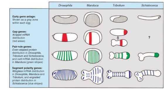
Much of what has been learned about the molecular process of segmentation is from Drosophila; how much is representative of a general process for all insects is not yet known. The segment polarity and homeotic genes, as well as their presumed functions, seem to be conserved in all insects examined so far; however, the activity of the maternal coordinate, gap, and pair-rule genes is more variable (Fig. 4). Because of the variation in the formation of germ anlage, it is not surprising that the earliest stages of the segmentation gene cascade established in Drosophila do not function in more ancestral insects. Exactly how short-germ-type embryos establish their segmental pattern remains to be discovered.
THE GERM BAND AND DORSAL CLOSURE
The germ band is a two-layered structure, comprising both ectoderm and mesoderm that represents the outline of the final body plan along both axes. As the embryo grows, the germ band transforms from this essentially two-dimensional, two-layered sheet into a three-dimensional larva. From anterior to posterior, all the segments are represented. Individual segments first become visible near the anterior end, where the ectoderm differentiates into the brain and compound eyes. Protrusions develop anterior to the mouth opening that will eventually grow to form the labrum (front lip of mouth-parts) and the antennae. The next segment, the intercalary segment, develops a transient limb bud, which is later retracted. This bud may be a remnant of a second pair of antennae found in this position along the anterior-posterior axis in crustaceans. Each of the first three segments behind the mouth form paired appendages that become the mouthparts: mandibles, maxillae, and labium. The next three segments develop into the thorax and form appendages that become walking legs. During the remainder of embryogenesis, as organs develop and differentiate, the flanks of the germ band, both ectoderm and mesoderm, grow laterally and extend around the yolk. The two edges of the germ band meet and fuse along the dorsal mid-line, such that the mesodermal and ectodermal layers now enclose the yolk.
ORGANOGENESIS
When the germ band is fully segmented and gastrulation is complete, the remainder of embryogenesis involves the differentiation of the ectoderm and mesoderm into the organ systems of the larva or juvenile. The ectoderm gives rise to the bulk of the larval or adult form. Most obviously the ectoderm forms the “skin” of the larvae, marked by numerous bristles and hairs. In addition, the nervous system develops from the ventral ectoderm, and the tracheal system develops from invaginations of the lateral ectoderm. Ocelli, salivary glands, a prothoracic gland, corpora allata, molting glands, oenocytes, and silk glands also develop as ectodermal invaginations. Finally, two additional invaginations of the ectoderm occur:
1. The stomadeum occurs in a central position near the anterior of the germ band, and once invaginated, these cells proliferate in a posterior direction to form the foregut.

FIGURE 4 Comparative germ band and expression patterns of segmentation genes.
2. The proctodeal invagination occurs in the terminal segment, and these cells grow anteriorly to form the hindgut.
Malpighian tubules, the insect excretory organ, develop from out-pocketings of the proctodeum. The invaginated mesoderm initially forms a pair of transient coelomic sacs in each segment (Fig. 1E). From these, the dorsal vessel, or heart, the internal reproductive organs, muscles, fat body, subesophageal gland, and hemocytes will form. The midgut arises from a third germ layer, the endoderm, that develops at the edge of the fore- and hindgut invaginations and eventually fuses with them to complete the gut. During the remainder of development both the mesodermal and ectodermal organ pri-mordia all undergo differentiation into tissue-specific cell types and cell rearrangements required to form the final organ structures.
APPENDAGE DEVELOPMENT
In direct-developing hemimetabolous insects, leg and wings develop as direct outpocketings from the lateral embryonic ectoderm. Leg buds appear early, just after the completion of gastru-lation, whereas wing buds appear later in development, after the lateral ectoderm has grown dorsally. In many metamorphic insects, rather than outpocketing, a cluster of cells that will form the adult leg and wing invaginates below the ectoderm. These cells become the leg and wing imaginal discs and do not undergo any further differentiation until later larval stages.
The molecular basis of positioning the limb primordia within the embryo is also well established in Drosophila and seems to be similar in many respects throughout both hemi- and holometabolous insects. The same information required to pattern the body axis (Fig. 2) is used to pattern the limb primordia. Every segment has the capacity to form a limb, and limbs appear at a discrete boundary formed at the intersection of the segment polarity genes, and the graded signals that are used to pattern the dorsal ventral axis of the embryo. Limb primordia are marked by the expression of the Distal-less gene. The absence of Distal-less gene function results in the loss of distal limb structures up to the proximal limb segment, which is the coxa. The limbless abdomen characteristic of insects is created by a subsequent repression of Distal-less gene activity by several of the homeotic genes.
HATCHING
Upon completion of organ formation and histogenesis, the embryo begins to stretch and contract its newly formed muscles, and gas is secreted into the trachea. Embryogenesis is over when the maternally supplied yolk has been consumed. Hatching is achieved by any number of means, but typically, hatching is a mechanical process, in which the larva either chews its way out of the chorion, grows by imbibing air until the chorion cracks, or uses a special egg burster. There is sometimes an enzymatic digestion of the eggshell, but complicated hydrostatic mechanisms are used, as well. The hatchling emerges as a first instar (larva or nymph).
