Vesiculobullous diseases, which number more than 50, are characterized by fluid-filled blisters in the skin. Blisters smaller than 0.5.cm are called vesicles, and larger ones are called bullae. Vesicles and bullae are reaction patterns of skin to injury and thus can be caused by a wide variety of conditions.
Most primary vesiculobullous diseases are either immunolog-ic or genetic. They are caused by autoimmune reactions to components of skin, by allergic reactions to external agents in which the skin is the major organ system affected, and by genetic conditions in which some components of the skin are missing or abnormal. The final common pathway is disadhesion: one or more of the structures that hold the skin together separate, and a fluid-filled cavity appears. The different diseases are classified by the structure or structures affected and the mechanism or mechanisms by which disadhesion occurs [see Table 1]. In this subsection, several paradigmatic vesiculobullous diseases are discussed in the context of a general diagnostic approach to the patient with blistering lesions.
General Clinical Assessment
Diagnosis is based on clinical features, histologic findings, and immunologic findings. Clinical features of diagnostic importance include the following:
1. The history. Is the condition acute or chronic? Is it aggravated by sun or physical trauma?
2. The appearance of individual lesions [see Table 2 ]. Is the lesion a vesicle or bulla? Is it tense, flaccid, or umbilicated? Does the skin at the base of the blister appear normal, urticarial, or scarred? Is the border of each urticarial lesion annular or oval or is it irregular? Is the blister in the middle of urticarial plaques or on the periphery? Do more than one bulla arise from the same plaque?
3. The grouping of individual lesions. Are the lesions in closely spaced groups (as occurs in herpes simplex), or are they randomly distributed?
4. Sites of involvement. Are lesions on mucosal surfaces as well as on the skin? Are they predominantly on flexural or extensor surfaces; on the palms and soles or on the dorsa of the hands and feet; on the scalp, face, and upper torso; or on areas exposed to trauma?
The most important histologic finding is the layer of skin where the blister forms. If the blister forms in the epidermis, does it form immediately above the basal cell layer or higher up (beneath the stratum corneum)? If it forms in the basement membrane zone, is it within the lamina lucida or below the lamina densa? The precise location may be determined by immuno-fluorescence or by electron microscopic procedures.
The most important immunologic finding is the presence or absence of abnormal circulating or tissue-fixed antibodies to skin. These are usually detected by immunofluorescence techniques: (1) indirect immunofluorescence to detect circulating antibodies and (2) direct immunofluorescence on skin biopsy specimens to detect tissue-fixed antibodies. Recently, enzyme-linked immunosorbent assays (ELISAs) using purified antigens have become available to detect the antibodies that occur in some of the bullous diseases, such as pemphigus.
Pemphigus
Definition and pathogenesis
Pemphigus is characterized by blisters that arise within the epidermis and by a loss of cohesion of the epidermal cells (acan-tholysis) that results in the formation of clefts above the basal cell layer. Autoantibodies directed against adhesion molecules cause epidermal keratinocytes to separate, resulting in intraepidermal bullae. There are two types of pemphigus: deep (e.g., pemphigus vulgaris) and superficial (e.g., pemphigus foliaceus). They differ in the epidermal layers that are injured, in the clinical manifestations of the diseases, and in the associated immunologic abnor-malities.1 In the deep forms, the blisters form immediately above the basal cell layer and are associated with autoantibodies to desmoglein 3; about half the cases are associated with antibodies to desmoglein 1 glycoprotein keratinocyte adhesion molecules.2 In the superficial forms, the bullae form immediately below the stratum corneum. The superficial forms of pemphigus are associated with antibodies to desmoglein 1.
Clinical features
Pemphigus Vulgaris
Pemphigus vulgaris is the most common form of pemphigus. It can develop at any age but usually occurs in persons between 30 and 60 years old. The disorder tends to affect persons of Mediterranean ancestry but can occur in persons of any ethnicity. Pemphigus is more common in persons with certain HLA allotypes. The occurrence of the disease in first-degree relatives, although rare, suggests an inherited susceptibility transferred as a dominant trait. However, other unknown factors are required for expression of the disorder in predisposed persons.3 Studies of HLA class II alleles in Japanese patients as well as in other ethnic groups show an association with HLA-DRB1*04 and HLA-DRB1*14 in patients with pemphigus vul-garis across racial lines.
Pemphigus vulgaris usually, but not invariably, begins with chronic, painful, nonhealing ulcerations in the oral cavity [see Figure 1 ]. Bullae are rarely seen because they rupture easily, leaving ulcerated bases. The ulcerations are usually multiple, superficial, and irregular in shape. Any oral mucosal surface can be involved, but the most common sites are the buccal and labial mu-cosae, the palate, and the gingiva. The occurrence of multiple ulcerations differentiates these lesions from ulcerated malignant tumors of the oral cavity, which are usually single. A diagnosis of pemphigus is usually considered only after lesions have been present for weeks to months.
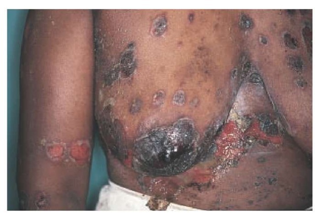
Skin lesions can also be the initial manifestation, beginning as small fluid-filled bullae on otherwise normal-looking skin. The blisters are usually flaccid because the thin overlying epidermis cannot sustain much pressure. Bullae therefore rupture rapidly, usually in several days, and may be absent when a patient is examined. Sharply outlined, coin-sized, superficial erosions with a collarette of loose epidermis around the periphery of the erosions may appear instead. The upper chest, back,scalp, and face are common sites of involvement, but lesions can occur on any part of the body. The condition progresses over weeks to months [see Figure 2]. Sites often overlooked include the periungual areas (manifested as painful, erythema-tous, paronychial swelling), the pharynx and larynx (pain on swallowing and hoarseness), and the nasal cavity (nasal congestion and a bloody mucous discharge, particularly noticeable upon blowing the nose in the morning).
Table 1 Differentiating Features and Standard Therapy for Selected Blistering Diseases
BMZ—basal membrane zone
IC—intercellular
IF—immunofluorescence
IVIg—intravenous immunoglobulin
Table 2 Pathologic Typology of Blisters57
|
Blister Type |
Mode of Formation |
Site of Formation |
Disease |
|
|
Subcorneal blister |
Detachment of horny layer |
Epidermis (subcorneal layer) |
Miliaria crystallina Impetigo |
|
|
Blister due to intracellular degeneration |
Separation of cells from one another |
Upper epidermis |
Friction blisters |
|
|
Spongiotic blister |
Intercellular edema |
Epidermis |
Dermatitis (eczema) Miliaria rubra |
|
|
Acantholytic blister |
Dissolution of intercellular bridges |
Epidermis (suprabasal layer) |
Keratosis follicularis (Darier disease) Pemphigus vulgaris |
|
|
Epidermis (subcorneal layer) |
Pemphigus foliaceus |
|||
|
Viral blister |
Ballooning degeneration leading to acantholysis |
Epidermis |
Herpes simplex Herpes zoster Varicella |
|
|
Blister due to degeneration |
Cytolysis of basal cells |
Basal cell layer |
Epidermolysis bullosa simplex Erythema multiforme (epidermal type) |
|
|
of basal cells |
Loss of dermal contact by damaged basal cells |
Basal cell layer |
Lichen planus Lupus erythematosus |
|
|
Blister due to degeneration of basement membrane zone |
Damage in the structures that cause coherence of basal cells |
Basement membrane zone |
Bullous pemphigoid Dermatitis herpetiformis Erythema multiforme (dermal type) |
|
|
Dermolytic blister |
Anchoring fibrils are decreased and rudimentary |
Dermis |
Dystrophic epidermolysis bullosa Acquired epidermolysis bullosa |
A characteristic feature of all severe active forms of pemphigus is the Nikolsky sign, in which sliding firm pressure on normal-appearing skin causes the epidermis to separate from the dermis. The Nikolsky sign is elicited most easily on clinically un-involved skin adjacent to an active lesion.
If left untreated, the erosions and bullae of pemphigus vul-garis gradually spread, involving an increasing surface area, and can become complicated by severe infections and metabolic disturbances. Before the advent of corticosteroids, pemphigus was almost invariably fatal—approximately 75% of patients died within a year.5 However, as better techniques have permitted the diagnosis of earlier, milder forms of the disease, the prognosis has improved significantly.6 Mild forms may regress spontaneously, and the progression of even the most severe forms can be reversed in most cases. With treatment (see below), lesions normally heal without scarring. Most patients treated for pemphigus will enter a partial remission within 2 to 5 years. They can then be maintained lesion-free with minimal doses of corticosteroids (approximately 15 mg of prednisone daily). In a longitudinal study of outcome in 40 patients with pemphigus vulgaris, 45% entered a complete and long-term remission after 5 years and 71% after 10 years. Patients in remission remained lesion-free without any therapy.7 The hyperpig-mentation that is commonly associated with pemphigus usually resolves after several months.
In pregnancy, pemphigus appears to be associated with an increased incidence of premature delivery and fetal death.8 The lesions of pemphigus can appear on the skin of the neonate; however, they normally resolve spontaneously in several weeks.
Pemphigus Foliaceus
Pemphigus foliaceus is the second most common form of pemphigus. It usually begins with small (approximately 1 cm), pruritic, crusted lesions resembling corn flakes on the upper torso and face. The crusts are easily removed, leaving chronic, superficial erosions.
Over weeks to months, the condition progresses, with an increasing number of lesions appearing on the upper torso, face, and scalp. In extensive cases, lesions develop over the entire body, become confluent, and can progress to an exfoliative eryth-roderma. In contrast to the deep forms of pemphigus, oral involvement in pemphigus foliaceus is very rare.
The prognosis of untreated pemphigus foliaceus is more favorable than that of pemphigus vulgaris. The lesions of pemphi-gus foliaceus are not as deep, and there is less chance for infection, fluid loss, and metabolic disturbance.
Figure 1 Painful ulcerations or erosions in the mouth may be present many months before the onset of generalized pemphigus vulgaris.
Figure 2 Flaccid bullae of pemphigus vulgaris have broken down to form erosions and crusts, particularly under the breasts.
Although pemphigus foliaceus is less severe, the doses of medications required for control are similar to those used for pemphigus vulgaris. There are two clinical variants: pemphigus erythematosus and fogo selvagem. Pemphigus erythematosus (also known as Senear-Usher syndrome) has features of lupus erythematosus. Fogo sel-vagem (Portuguese for "wild fire"; also known as endemic pemphigus and Brazilian pemphigus) [see Table 1] may be triggered by exposure to one or more environmental antigens.


