Image Processing Reference
In-Depth Information
(3)
When the cases in a node are evenly distributed across the categories, the Gini index takes
its maximum value of 1−(1/
k
), where
k
is the number of categories for the target atribute. Fur-
thermore, for all cases in the node that belong to the same category, the Gini index equals zero.
6 The proposed algorithm
The goal of this chapter is to describe a process for detecting and distinguishing between be-
nign and distorted blood cells in a colored microscopic image. This work can help doctors,
physicians, chemists, and anyone else who cares about blood cell detection and analysis, and
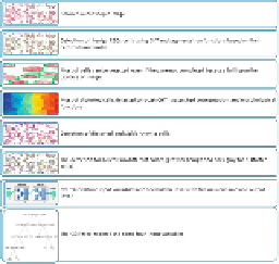
determination of diseases. The proposed algorithm is divided into two steps, as shown in
Fig-
ure 5
.
FIGURE 5
The main points of the proposed algorithm.
In the first step, I apply a CHT with morphological functions to the bright and dark intensity
of cells to detect and count benign and distorted blood cells. In the second step, I use NN and
a C&R tree to test and check the performance of the proposed algorithm for diagnosing a pa-
tient with sickle-cell anemia, evaluating which process is more effective than the other. Clas-
siication and prediction depend on the detected cells' data variables (e.g., area, convex area,
perimeter, and eccentricity) to reduce the errors in detection operation and ensure final dia-
gnosis of sickle-cell anemia. The first part of the CHT includes many operations:
•
Cell polarity
indicates whether the circulating blood cells are brighter or darker than the
background.
•
Computation
(two-stage) calculates the accumulator array of the CHT. It is based on com-
puting radial histograms; radii are clearly applying the estimated cell centers along with
the image information [
20
].
• The
sensitivity factor
is the sensor of the accumulator array in the CHT. Detection includes
weak and partially hidden or overlapped cells; however, higher sensitivity values increase
the risk of false detection.
This stage also determines the edge gradient threshold, given that cells generally have a
darker interior (nuclei) surrounded by a bright halo. The edge gradient threshold is very use-
ful for determining edge pixels, and both unhealthy and strong blood cells can be detected

Search WWH ::

Custom Search