Chemistry Reference
In-Depth Information
“bulk” measurements is the lack of easy integration of a triggered release of
Ca
2
+
.
Therefore, in conjunction with experiments performed in a cuvette, a microfluidic
technique was developed for the investigation of the
Ca
2
+
triggered SNARE fusion.
Microfluidic techniques offer the advantage of reduced sample volume and con-
trolled mixing of fluids. Microfluidic devices are made using standard
photolithographic techniques as described in the appendix. Two CCD cameras with
appropriate optical filters were integrated with an inverted microscope (Olympus,
IX81) as shown in Fig.
3.4
as a means to measure FRET efficiency. A microfluidic
device is mounted on an X-Y stage and the donor fluorescence is excited by the
appropriate use of an optical filter (480nm) and the resulting images from the donor
(540nm) and acceptor (610nm) fluorescence are simultaneously recorded in two
separate CCD cameras (PCO 1200).
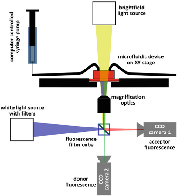
Fig. 3.4
Experimental setup for performing microfluidic FRET experiments to study membrane
fusion. A microfluidic device made using PDMS soft lithography is mounted on an inverted micro-
scope. The flow through the device is regulated by computer controlled syringe pumps. The FRET
fluorescence is recorded on two seperate CCD cameras, one for the donor and the other for the
acceptor fluorescence wavelengths respectively. The donor fluorescence is excited by using filtered
light from a white light source