Biomedical Engineering Reference
In-Depth Information
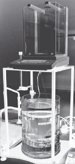
Transducer
attachment
Balance
Water bath
Oil target
FIGURE 5.12
Buoyancy type system for making ultrasound output measurements.
field (Kudo et al. 2009). Sending light through the ultrasound
field onto a screen or detector produces a form of shadow, which
reveals the overall shape of the field. Scanning laser vibrometry
can be used in a similar way (Zipser and Franke 2004). An alterna-
tive approach is to look at the thermal effect of an acoustic field on
a known medium, as described in the following section.
used to monitor the consistency of the output of transducers, the
main important consideration is that measurements made over
time are comparable. However, when relating quantities such as
pressure and intensity to safety indices, it is necessary to account
for attenuation of the acoustic field as it passes through the body.
This is known as derating. The nominal quoted value for the
attenuation of ultrasound by soft tissues in the AIUM/NEMA
standard (AIUM and NEMA 1992) is 0.3 dB cm
−1
MHz
−1
. A
known pressure or intensity at a point in water can be derated
to provide the corresponding value at a particular depth in the
body, provided the frequency of the ultrasound beam is known.
It is well documented that different tissue types have differ-
ent ultrasound attenuation coefficients, as discussed in Section
5.4. Although having a single accepted value for the approximate
attenuation coefficient in the body is useful for giving an esti-
mate of the peak intensities and pressures incident on tissues,
this one value is not representative of all scenarios, and inac-
curate assumptions have the potential to affect treatment out-
comes. It is therefore important to interpret such values with
caution and make measurements of attenuation coefficients of
exposed media or of in situ pressures where practicable.
5.5.3.5 thermal Calibration
A relatively quick and simple method of assessing transducer
output is to look at the temperature rise produced in a well-char-
acterized medium. Absorbing devices have been produced that
rely on thermochromic materials that change color or hue with
temperature. These display a reversible change for temperatures
appropriate for physiotherapy ultrasound, and can be quantified
by photographing the effects and calibrating the color change
through image processing (Martin and Fernandez 1997, Shaw
et al. 1999). More recently, a pyroelectric device based on an
absorbing piezoelectric material with a voltage output relating
to temperature rise in the material has been created, as described
previously in relation to power measurement (Zeqiri and Barrie
2008). Although these give a measure that relates to the temporal
average intensity of an ultrasound field, it is important to make
peak pressure and intensity measurements, particularly of HIFU
fields, in order to fully understand the potential bioeffects.
5.5.3.7 Safety Indices
Thermal and mechanical indices have been defined and dis-
cussed briefly in Section 5.4. Equation 5.20 shows how the TIS
can be calculated if the output power has been measured as
described in the previous paragraphs, and if the center frequency,
f
c
, of the acoustic wave is known. The latter can be found using
5.5.3.6 Derating
Calibration measurements for HIFU devices are typically made
under free field conditions, for example, in a large water tank
where any field perturbations are minimized. As QA data are