Biomedical Engineering Reference
In-Depth Information
350
100
∆
R
0
300
−
100
250
−
200
200
−
300
150
400
−
∆
f
100
−
500
50
−
600
0
−
700
−
800
−
50
0
5
10
15
20
25
Time (h)
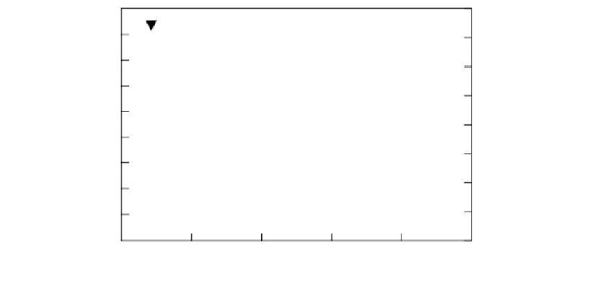
FIGURE 1.30
Kinetics of
R
shifts produced in a QCM biosensor during its formation by the addition of 50,000 ECs at
the time indicated by the arrowhead position. Prior to cell addition, media and serum-lacking cells were incu-
bated for 2 h to establish baseline
f
and
R
values for calculation of the
f
and
R
shift values from the differences
at all times following cell addition. Reprinted with permission from Marx, K.A., Zhou, T., Warren, M., Braunhut,
S. J. (2003). Quartz Crystal Microbalance Study of Endothelial Cell Number Dependent Differences in Initial
Adhesion and Steady-State Behavior: Evidence for Cell-Cell Cooperativity in Initial Adhesion and Spreading.
Biotechnol. Prog
. 19:987-999. Copyright (2003) American Chemical Society.
f
and
monitoring tool to study the time-dependent surface attachment of ECs on the gold QCM
surface. This involved first determining the requirements for cell attachment and main-
tenance in growth media and serum in a sterile environment. Then we were able to
determine the time course required to achieve steady-state attachment values of the
measure
R
shift parameters. Also, we determined that with increasing cell num-
ber added, the magnitude of these QCM shift parameters were well correlated with the
number of electronically counted cells released following trypsinization from the QCM
surface (93,94). In this way, the biosensor accurately reflects the population of ECs, up to
about 20,000/0.196 cm
2
, stably attached to the QCM surface. We demonstrated that dur-
ing their transition from initial contact and adhesion to stable attachment, cells exhibited
increasing amounts of energy dissipation as expressed through increasing levels of
motional resistance measured for the QCM crystal. Viewed as a progression of time
points in Figure 1.31 (following the arrows), the frequency and resistance shift data for
cell attachment showed that the cells at the surface initially exhibited pure liquid behav-
ior [(
f
and
)
1/2
dependence] following the linear fit to the pure sucrose measured data. With
increasing time, the data points evolve to indicate that motional resistance and energy
dissipation properties at the crystal surface have increased significantly, to values char-
acteristic of the steady state of the cells. These properties are consistent with the follow-
ing known qualitative picture of anchorage-dependent normal cell attachment behavior.
In the cell attachment process, an ECM of specific proteins is secreted by the cell upon a
surface. Integral membrane protein receptors on the cell surface termed integrins then
recognize specific peptide sites within ECM and a stable attachment is formed. The inter-
nal protein cytoskeleton of the attached cell, using these external integrin anchor points
to ECM, then forms a networklike structure that maintains the spread shape of the
attached cell upon the matrix surface. The cell's internal protein structure responsible for
the spread shape has been termed the tensegrity structure (95). Tensegrity structures
formed within the cells are consistent with the significantly increased motional resistance