Biomedical Engineering Reference
In-Depth Information
20
16
20
12
16
12
8
8
4
4
0
0
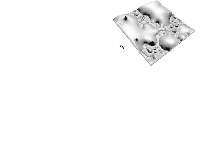
FIGURE 1.29
(See color insert)
Noncontact AFM three-dimen-
sional views of a 1:1 comonomer mixture of DELT
(above cmc) and
L
-tyrosineamide prior to (upper
image) and following (lower image) enzymatic poly-
merization with HRP and subsequent 24-h immer-
sion of a gold-coated mica substrate to immobilize
the complexes upon. Reprinted with permission
from Marx, K.A., Lee, J.S., Sung, C. (2004). Enzymatic
Copolymerization Alters the Structure of
Unpolymerized Mixtures of the Biomimetic
Monomers: The Amphiphilic Decyl Ester of
L
-
Tyrosine and
L
-Tyrosineamide—An AFM
Investigation of Nano- to Micrometer Scale Structure
Differences.
Biomacromolecules
5:1869-1876.
Copyright (2004) American Chemical Society.
5
4
5
3
4
3
2
2
1
1
0
0
of the long-fibrillar aggregates as the reaction product was being formed. This QCM
biosensor approach has the potential for application to many other experimental situa-
tions where optical techniques cannot be employed for sensing.
1.2.3.2 Quartz Crystal Microbalance Cell Biosensor for Cell Characterization and Drug
Discovery Applications
With the advent of solution-based QCM devices in the 1980s, investigators began to apply
the technology sporadically to studies of living cells. Two types of studies were per-
formed. The first was aimed at understanding fundamental cellular processes, and the sec-
ond was the use of the QCM as a cellular biosensor of some analyte. A number of recent
comprehensive reviews, including one from this Center, have described this cell-oriented
research area (65,89-91). At the Center for Intelligent Biomaterials, we have carried out
studies of both fundamental cellular processes as well as the integration of cells into QCM
biosensors. Our interest in the creation and characterization of cell QCM biosensors was
based upon our desire to exploit the wide range of intelligent properties possessed by
living cells. The current end products of evolution, cells, are exquisitely complex biological
elements that await more widespread integration into biosensors of varying design.
1.2.3.2.1 Measuring the Fundamental Process of Cell Attachment During Biosensor
Formation
The sensitive mass detection and surface motional resistance measurement capability of
the QCM signal transduction platform provides a unique capability for illuminating the
process of cellular attachment to surfaces. During the development of our cell QCM
biosensor, we studied the behavior of normal ECs during their attachment to the gold
upper electrode surface of the QCM platform (92,93). As Figure 1.30 illustrates, we first
demonstrated that the QCM device could be used as a sensitive and continuous