Biomedical Engineering Reference
In-Depth Information
350
24 h
300
Increasing energy
dissipation
250
200
43 min
150
100
50
Viscoelastic region
t
= 0
0
Pure elastic mass (sauerbrey Eq. 1)
−
50
−
700
−
600
−
500
−
400
−
300
−
200
−
100
0
100
∆
f
(Hz)
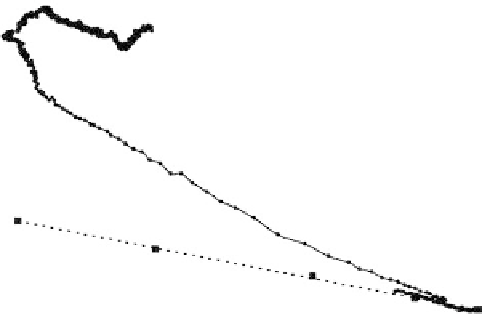
FIGURE 1.31
A
f
diagram of the time course following addition of 30,000 ECs to the QCM surface. Arrows indicate the
time course of the data points starting at the lower right with the
t
= 0 point at the arrowhead. The final time
point occurs for the cells at 24 h at their steady state where maximum energy dissipation is exhibited. The
R
-
R
-
f
behavior of a pure elastic mass is indicated by the horizontal line. The
f
points (filled squares) determined
experimentally for a series of increasing concentration sucrose solutions are depicted with the best fit dashed
line. This is the behavior of a Newtonian fluid producing a pure (
R
-
)
1/2
density-viscosity effect on the QCM
f
values. Reprinted with permission from Zhou, T., Marx, K.A., Warren, M., Schulze, H., Braunhut, S. J.
(2000). The Quartz Crystal Microbalance as a Continuous Monitoring Tool for the Study of Endothelial Cell
Surface Attachment and Growth.
Biotechnol. Prog.
16:268-277. Copyright (2000) American Chemical Society.
R
-
and energy dissipation properties exhibited by the steady-state cells that we have
observed on the oscillating quartz crystal. It is noteworthy that the QCM biosensor is
detecting energy dissipation levels for cells reaching steady-state attachment in Figure
1.31 that are above the pure density-viscosity effect, (
)
1/2
, line. This is significantly
greater viscoelastic behavior than the energy dissipated by unpolymerized or even HRP-
polymerized fibrillike DEDT structures binding to the gold surface (Figure 1.27) that pro-
duced measured values lying below this (
)
1/2
line.
In a more detailed QCM study, we examined the normal EC attachment process as a
function of cell number added (93). We observed a short 10-min time lag in the attachment
process before cells were able to produce measurable QCM frequency and motional resist-
ance shifts. Also, the QCM provided evidence for the existence of novel cell-cell coopera-
tivity behavior in the initial stages of cell adhesion and spreading (93). This was
manifested in the Figure 1.32
R
shift data plotted against cell number trypsinized
from the QCM surface in any given experiment. This trypsinized cell number reflected an
accurate indication of the number of firmly attached cells. The curve shape best fit to the
data at 1 h postcell addition was sigmoid shaped. However, the 24 h postcell addition, or
steady-state condition, had a best-fit hyperbolic curve shape. The sigmoid curve shape
behavior at 1 h following cell addition is characteristic of a cooperative process. It likely is
caused by the dependence of cell attachment on a mechanism involving some type of
cell-cell communication that is number or density dependent. Once established at their
steady state upon the surface, we also studied EC growth stimulation by the effect of
adding fibroblast growth factor protein and observing cell growth continuously for up to
4 days (92).
f
and