Biomedical Engineering Reference
In-Depth Information
sciaticnervesofnewbornWistarratsandculturedinanF12/DMEM
medium for
in vitro
expansion. The PGA fibers with a diameter
of 15
μ
m were arranged longitudinally into a cord and were then
seeded with Schwann cells suspended in a culture medium and co-
cultured for one week. In adult Wistar rats, the sciatic nerve was
exposed and a 15 mm long defect was created and then bridged
with the cell-scaffold construct that was wrapped with a degrad-
able biomembrane in an experimental group or bridged with a syn-
genic nerve graft as a positive control and bridged with PGA fibers
alone or left unrepaired as a negative control. Engineered nerves
were harvested at three months posttransplantation for histology
andelectrophysiologicalevaluation.Theresultsshowedthatperiph-
eral nerve function was much better recovered in the experimen-
tal and positive control groups than in the negative control group.
The gained functions of the engineered nerve were demonstrated
by the response of rat extremities to pain and temperature stim-
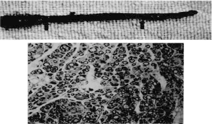
ulation and by the maintaining of gastrocnemius weight. Grossly,
the engineered nerve has a morphology similar to that of the nor-
mal peripheral nerve, whereas thePGA fiberswere totally degraded
in the negative control group. Electromyogram evaluation revealed
that no statistically significant difference was found between the
Figure 34.21.
Grossview(
top
,betweenarrows)andhistologyofatissue-
engineered nerve (
bottom
). (Reprintedby permission from Ref. 8).


Search WWH ::

Custom Search