Biomedical Engineering Reference
In-Depth Information
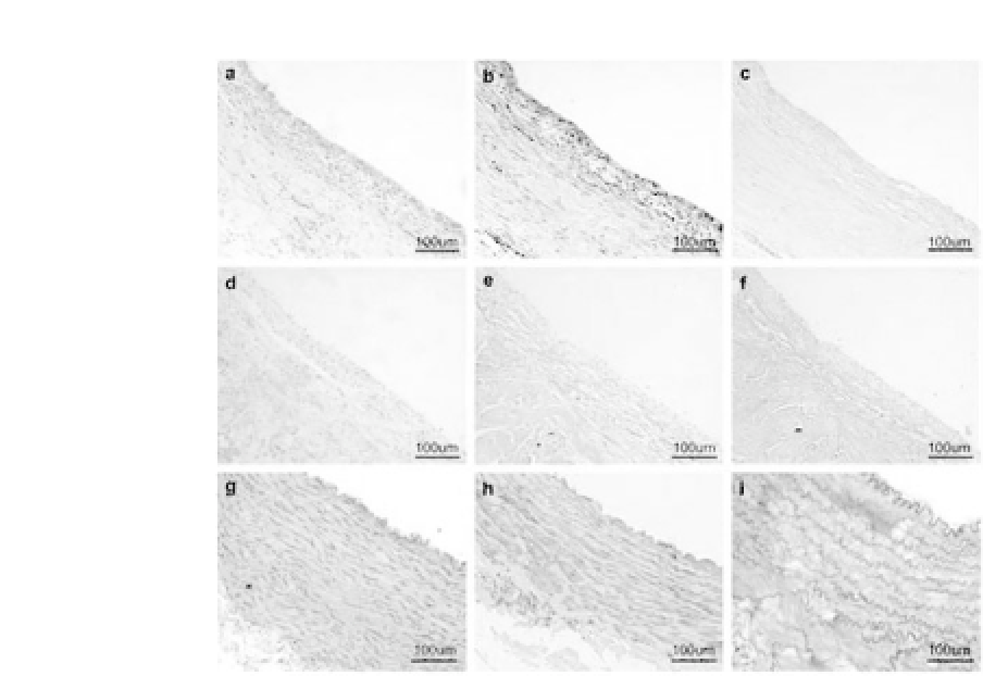
Figure 34.20.
Histology of the engineered vessel walls after 8 weeks of
culture with (a-c) or without (d-f) pulsatile stimulation. Native canine
abdominal arteries are presented as references (g-i). H&E staining shows
that PGA fibers are completely degraded and SMCs are in well-orientated
layers in the dynamic group (a), while SMCs are orientated randomly in the
static group (d). Masson staining shows well-organized collagenous fibers
distributed evenly in the vessel wall of the dynamic group (b), but only
disorganized collagenous fibers are present in the static group (e). Elastic
fibers are neither found in the dynamic group (c) nor in the static group (f)
by Gomori staining. Bars: 100
μ
m. (Reprinted by permission from Ref. 20).
See also Color Insert.
walls of the static group. In addition, mechanical properties such as
tensile strength, suture-holding retention strength, and burst pres-
sure were significantly enhanced in the dynamic group compared
withthecontrolgroup.Theseresultsindicatethatsuchanapproach
is suitable for engineering large-vessel wall tissue and for engineer-
ing other tissueswith amuscular tubularstructure.
20
34.7 PGA Fibers for Engineering Peripheral Nerve Tissue
PGA fibers were also investigated for their application in periph-
eral nerve engineering. Schwann cells were isolated from the


Search WWH ::

Custom Search