Biomedical Engineering Reference
In-Depth Information
(a)
(b)
(d)
(c)
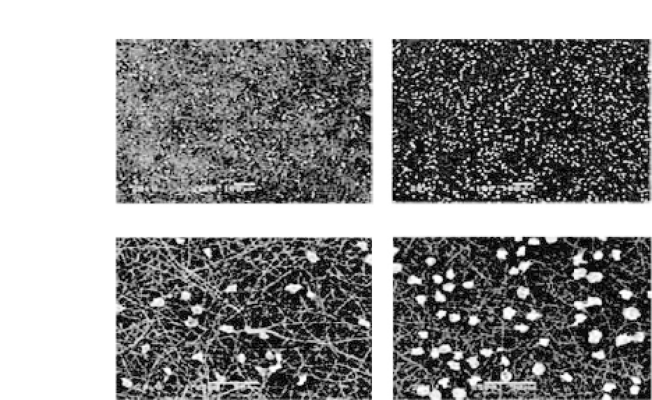
Figure 13.7.
SEM images of NIH3T3 cells cultured for 4 h on PHBV (a,b)
and PHBV-Col (c,d) nanofibrous scaffolds at an original magnification of
100
×
(a,c) and 500
×
(b,d) (adapted from Ref. 16).
scaffolds after a 4-hour culture. The cells adhered well on the sur-
facesofbothnanofibrousscaffolds.Morecellswereobservedonthe
surface of the PHBV-Col nanofibrous scaffold than that of PHBV.
47
Figure 13.8 shows the proliferation of cells on the nanofibrous scaf-
folds.CellproliferationonthePHBVnanofibrousscaffoldwassignif-
icantly accelerated by the incorporation of type I collagen (PHBV-
Col) (
P
<
0
.
01).
16
13.5.2
PHBV/Gelatin Nanocomposites
Cell culture experiments showed that NIH3T3 cells had very favor-
able interactions with the PHBV/gelatin composite scaffold com-
parison with the PHBV film and the PHBV nanofibrous scaffold.
Significantly, cellular infiltration into the PHBV/gelatin compos-
ite fibrous scaffold was demonstrated. It is concluded that co-
electrospinningtheECMwithsyntheticpolymers,suchasPHBV,has
many potential applications in tissue engineering.
4
,
8
As shown in
Fig. 13.9, NIH3T3 fibroblasts were highly dispersed on to the PHBV


Search WWH ::

Custom Search