Biomedical Engineering Reference
In-Depth Information
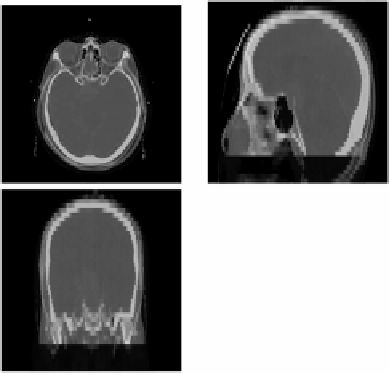
Figure 9.18:
The axial, sagital, and coronal views of the two CT brain volumes
(one in red, second one in green) prior to registration. Meaningful comparison
is difficult. (Color slide.)
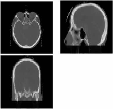
Figure 9.19: The axial, sagital, and coronal views of the two CT brain volumes
(one in red, second one in green) after the registration. The volumes are aligned,
and the large and medium-scale differences were compensated by the registra-
tion. This permits to identify more subtle differences. (Color slide.)