Biomedical Engineering Reference
In-Depth Information
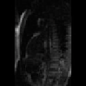
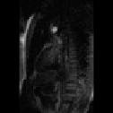
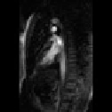
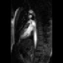
6
9
11
14
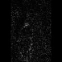
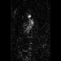
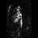
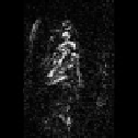
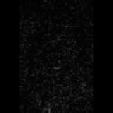
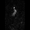
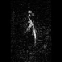
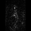
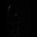
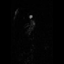
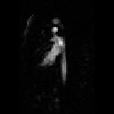
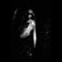
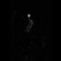
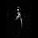
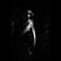
Figure 9.17: The first line presents original images number 6,9,11, and 14 from
a sequence of originally 60 images of myocardical perfusion MRI. The second line
presents the difference images between the original images and their immediate
predecessors; movement artifacts can be clearly seen. On the third line you
can see the difference images from the motion corrected sequence using our
algorithm; the movement artifacts are significantly reduced. The same effect
is also visible comparing the differences of the sequence images with the first
image of the sequence on the original (fourth line) and corrected (fifth line)
sequences.
This, together with the constant advances in computer technology will enable
truly interactive operation of automatic and semi-automatic elastic image reg-
istration with numerous applications in medicine, biology, and any other field
where deformed images need to be compared.