Biomedical Engineering Reference
In-Depth Information
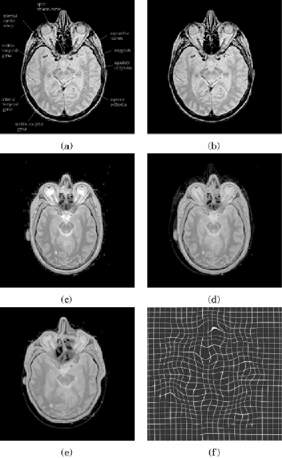
Figure 9.9: The reference MRI proton density brain slice from the atlas with (a)
and without labels (b). The sample test slice of a corresponding region (c). The
superposition (in red and green) of the two images before (d) and after the
registration (e). The deformation field (f). Cubic splines were used with knot
spacing of
h
=
32. The image size was 512
×
512 pixels. The difference between
images is only partially corrected by the unsupervised registration. Misalignment
of several structures is clearly visible. (Color slide.)