Biomedical Engineering Reference
In-Depth Information
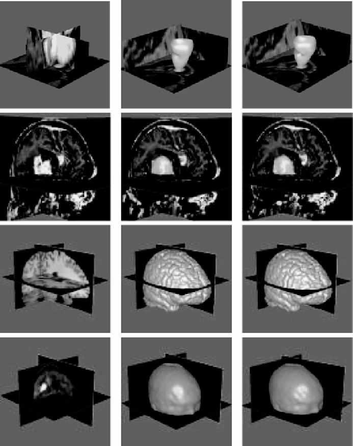
Figure 7.9: First row: A short-axis cardiac MR image and segmentation of
the left ventricular cavity. Second row: An MR brain image and segmentation
of the tumor. Third row: An MR brain image and segmentation of the brain.
Fourth row: A PET image and extraction of the surface of the head. The first
column shows the original images, the middle column shows the initial seg-
mentation results, and the right column shows the results after the necessary
modifications.