Biomedical Engineering Reference
In-Depth Information
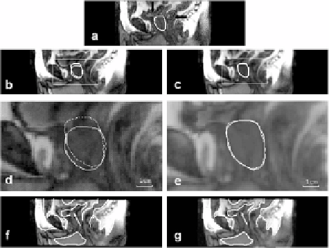
Figure 3.12:
Comparison of rigid body and warping registration for volumes
acquired with an interval of one week between imaging sessions. The reference
image (a) with a manually segmented prostate was acquired later with an empty
bladder (vertical arrow) and partial rectal filling (horizontal arrow). Images in
the left and right columns are from the floating volume acquired earlier following
rigid body and warping registration, respectively. To show potential mismatch,
contours from the reference are shown on images following registration, as
described in Fig. 3.11. The full bladder in (d) has pushed the prostate, shown by
the continuous curve, in the caudal direction. After warping, prostate contours
match closely (e). The bladder, rectum, and other organs closely align following
warping (g). With rigid body (f ), proceeding from left to right, the front of the
pelvis, the bladder (arrow), and the rectum are all misaligned. Images are sagittal
slices from S3.
(Fig. 3.12d). In addition, rigid body registration does not align the bladder and
parts of the rectum (Fig. 3.12f). With warping, the bladder closely matches the
reference, and the rectum is better aligned (Fig. 3.12g). Other visualization meth-
ods showed excellent alignment of internal and surface edges. Difference images
show that warping greatly improves alignment of internal structures as com-
pared to rigid body registration (Fig. 3.13). The difference image following rigid
body registration shows bright regions indicating misalignments (Fig. 3.13d)
that are removed with warping (Fig. 3.13e).
We also examined volume pairs with both volumes acquired in the diagnos-
tic position under comparable conditions. In the current data set, five volume