Biomedical Engineering Reference
In-Depth Information
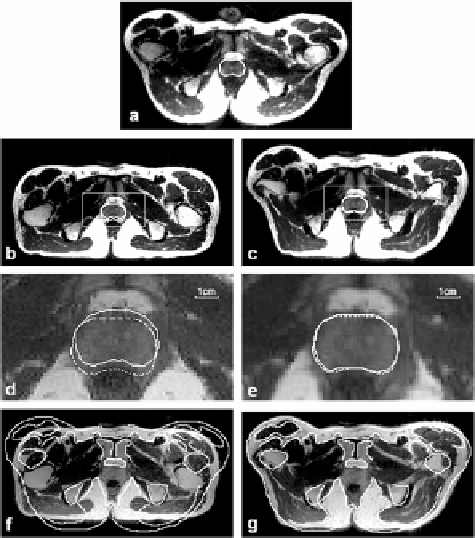
Figure 3.11: Comparison of warping and rigid body registration for volumes
acquired in the treatment and diagnostic positions. Image (a) is from the refer-
ence volume acquired in the treatment position, and the prostate is manually
segmented. Images in the left and right columns are from the floating volume
acquired in the diagnostic position following rigid body and warping registra-
tion, respectively. To show potential mismatch, the prostate contour from the
reference in (a) is copied to (b) and (c) and magnified as the dashed contours
in (d) and (e). The 3 mm movement of the prostate to the posterior is corrected
with warping (e) but not rigid body registration (d). Pelvic boundaries manually
segmented from the reference show significant misalignment with rigid body
(f ) that is greatly improved with warping (g). Images are transverse slices from
subject S2.
We next examine the effect of conditions such as bladder and rectal filling
that might change from one imaging session to the next. In Fig. 3.12 we compare
non-rigid and rigid body registration for a volume pair with one-week between
imaging sessions. One volume is with an empty bladder and the other is with
a relatively full bladder. There is also a difference in rectal filling. Non-rigid
registration closely aligns the prostate (Fig. 3.12e) while rigid body does not