Biomedical Engineering Reference
In-Depth Information
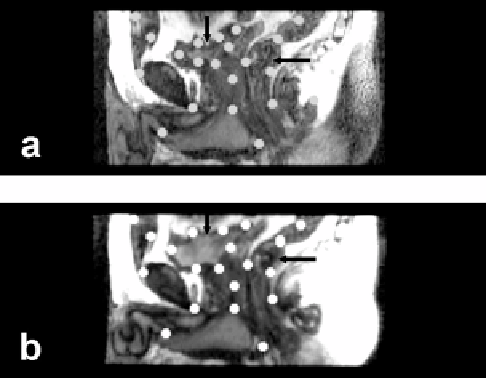
Figure 3.10:
Control point selection when images are acquired with a week's
interval between them. Image (a) is from the reference volume acquired one
week later with an empty bladder. Image (b) is to be warped and is from the
volume acquired earlier with a full bladder. Sagittal slices best show the defor-
mations at the bladder (vertical arrow) and rectum (horizontal arrow) where
most control points are placed. Volumes are from volunteer S3.
3.3.4.5
Registration Quality of Non-Rigid and Rigid
Body Registration
In Fig. 3.11 we compare non-rigid and rigid body registration for a typical volume
pair in the treatment and diagnostic positions. Following non-rigid registration,
the prostate boundary overlap is excellent (Fig. 3.11e) and probably within the
manual segmentation error. Similar results were obtained in other transverse
slices throughout the prostate. The prostate 3D centroid calculated from seg-
mented images displaced by only 0.6 mm, or 0.4 voxels, following warping.
Following rigid body registration, the prostate was misaligned with a displace-
ment to the posterior of
≈
3
.
4 mm when in the treatment position (Fig. 3.11d), as
previously reported by us [1]. Using rigid body registration, there is significant
misalignment throughout large regions in the pelvis (Fig. 3.11f ) that is greatly
reduced with warping (Fig. 3.11g). Note that warping even allows the outer sur-
faces to match well. Other visualization methods such as two-color overlays and
difference images, quickly show matching of structures without segmentation
but do not reproduce well on a printed page.