Biomedical Engineering Reference
In-Depth Information
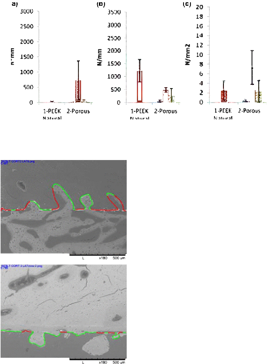
Figure 12.5
Showing the energy to failure (a), stiffness (b), and maximum shear (c) required for solid or porous PEEK
samples at 4 weeks for aged sheep (blue), 12 weeks for aged sheep (red), and 4 weeks for 18-month sheep (green).
machined topography) could have subtle effects on
the adhesion and mineralization of osteoblasts
[31]
;
therefore, the difference in topography and the
presence of pores may provide an added benefit for
some situations. The porous PEEK here had a mean
pore size of 70
m
m when analyzed using CT.
Although the mean pore diameter achievable through
this technology was lower than the majority of
recommendations for osseoconductivity in other
materials
[33
e
35]
, the smaller diameter here proved
sufficient to confer some benefit. Although larger
pore sizes seem to be preferred for the current
orthopedic porous biomaterials (e.g., porous
tantalum or titanium) and has been cited as needing
to be larger than 50 to 150
m
m
[36]
, the smaller pore
sizes are present in other application areas such as
CMF (e.g., porous polyethylene). It has been reported
that this is sufficiently effective for fibrous and bone
ingrowth in facial implants
[4,5]
. This may not be
optimal for ingrowth into a hydrophobic polymer and
the material may not possess the mechanical prop-
erties for other applications but it does suggest that
porosity, whatever the size, confers some benefits.
Some researchers have suggested that the necessary
pore size may only be required to be sufficient for
vascularization of the tissue to occur since this then
allows the subsequent nutrients to be present to
permit tissue ingrowth
[37]
. The interconnectivity of
this porous PEEK material was sufficient to permit
some limited ingrowth at the surface interface, but
the bulk of the material possessed closed cells, which
(a)
(b)
Figure 12.6
Showing the determination of bone
ongrowth areas using MatLab analysis of radiographs.