Biology Reference
In-Depth Information
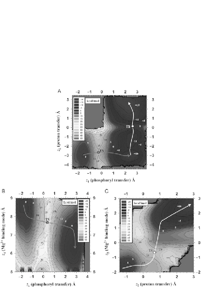
Mg
2
þ
-binding mode, and confirmed the sensitivity of the barriers to the
Mg
2
þ
ion position along the reaction coordinate. A common feature of
the reaction mechanism derived from the 3D profile was that the phosphoryl
transfer and general acid steps were stepwise, demonstrated in
Fig. 2.5
A,
allowing these steps to be decoupled.
Since both the phosphoryl transfer and general acid steps of the reaction
were coupled with Mg
2
þ
-binding mode, two separate 2D profiles were
generated for each step with a reaction coordinate corresponding to the
Figure 2.5 (A) Selected 2D surface in 3D free-energy profile simulations, harmonically
restrained along the coarse-grained metal ion-binding coordinate at d(Mg, G8:O2
0
)
¼
2.5 Å, where z
1
¼d(C1.1:O5
0
,P) d(P, G8:O2
0
), z
2
¼d(G8:O2
0
, G8:Ho2
0
) d(G8:Ho2
0
,
C1.1:O5
0
). (B) 2D PMF for Mg
2þ
-binding mode in phosphoryl transfer step, where
z
4
¼d(Mg, O5
0
)þd(Mg, G8:O2
0
). (C) 2D PMF for Mg
2þ
-binding mode in general acid step,
where z
5
¼d(Mg,O5
0
) d(Mg,G8:O2
0
). d(x, y) denotes distance between x and y.TSisthe
acronym of transition state.
Search WWH ::

Custom Search